
Surface and Radiological Anatomy — MCQs

On this page
The pleural reflection on the left midaxillary line is located in which intercostal space?
To which rib does the pleura extend in the mid-axillary line?
At which vertebral level is the fundus of the gallbladder typically located?
The tympanic note on percussion in Traube's space on the chest wall is due to which underlying structure?
What is the surface marking of the mitral valve?
Which of the following structures does the ureter cross on an abdominal radiograph?
What is the anatomical landmark for the mid-inguinal point?
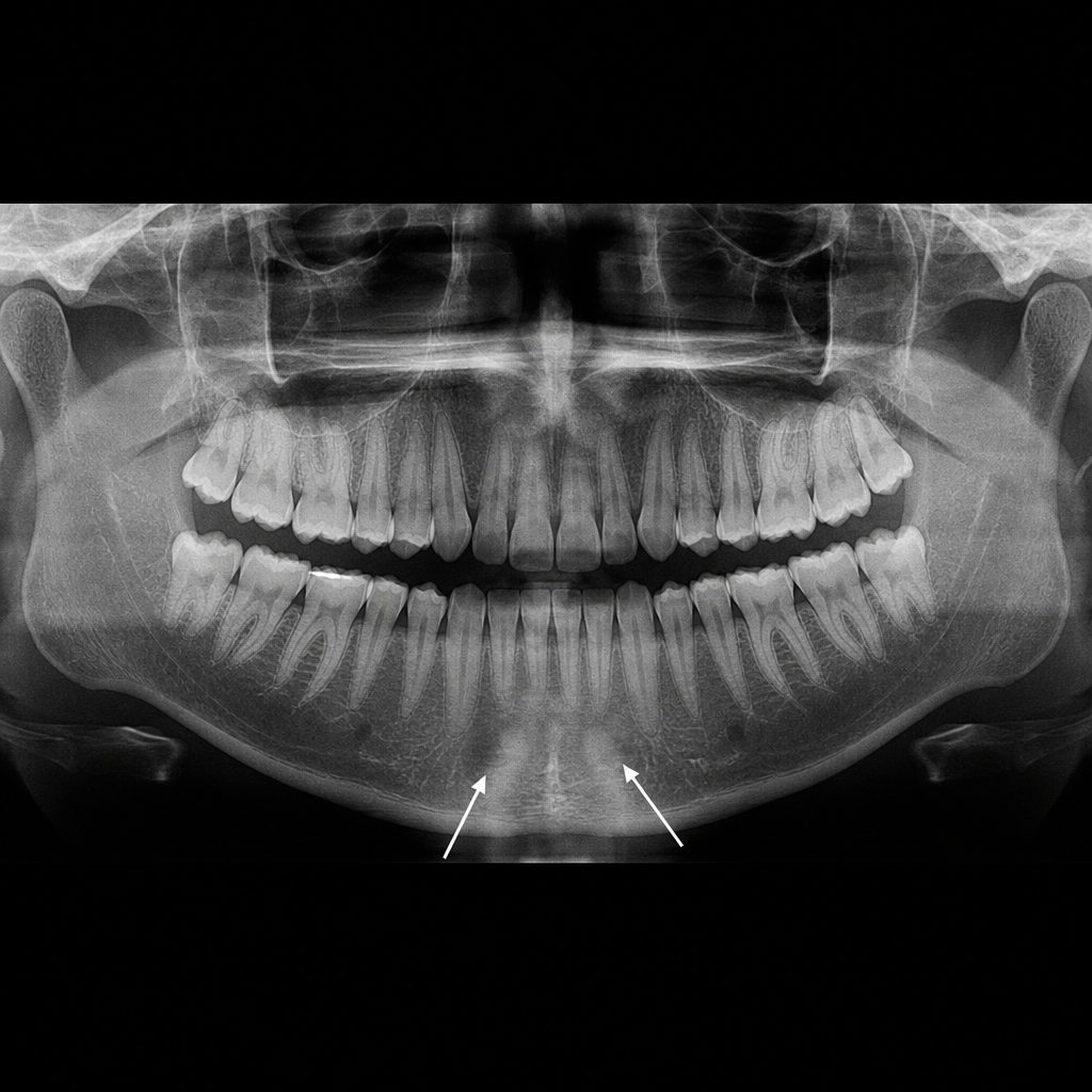
The symmetrical radiopacities marked with arrows are most likely?
The spine of the scapula can be palpated at which of the following vertebral level?
Identify the tarsal bone marked by the arrow in the given X-ray of the right foot.
Practice by Chapter
Surface Landmarks of the Head and Neck
Practice Questions
Surface Landmarks of the Thorax
Practice Questions
Surface Landmarks of the Abdomen
Practice Questions
Surface Landmarks of the Limbs
Practice Questions
Radiographic Anatomy
Practice Questions
CT Anatomy
Practice Questions
MRI Anatomy
Practice Questions
Ultrasonographic Anatomy
Practice Questions
Angiographic Anatomy
Practice Questions
Sectional and Cross-sectional Anatomy
Practice Questions
Anatomical Correlations in Common Imaging
Practice Questions
Interventional Radiological Anatomy
Practice Questions
Want unlimited practice?
Get full access to all questions, explanations, and performance tracking.
Scan to download app