Which anatomical structure classically displays mucin-producing glands?
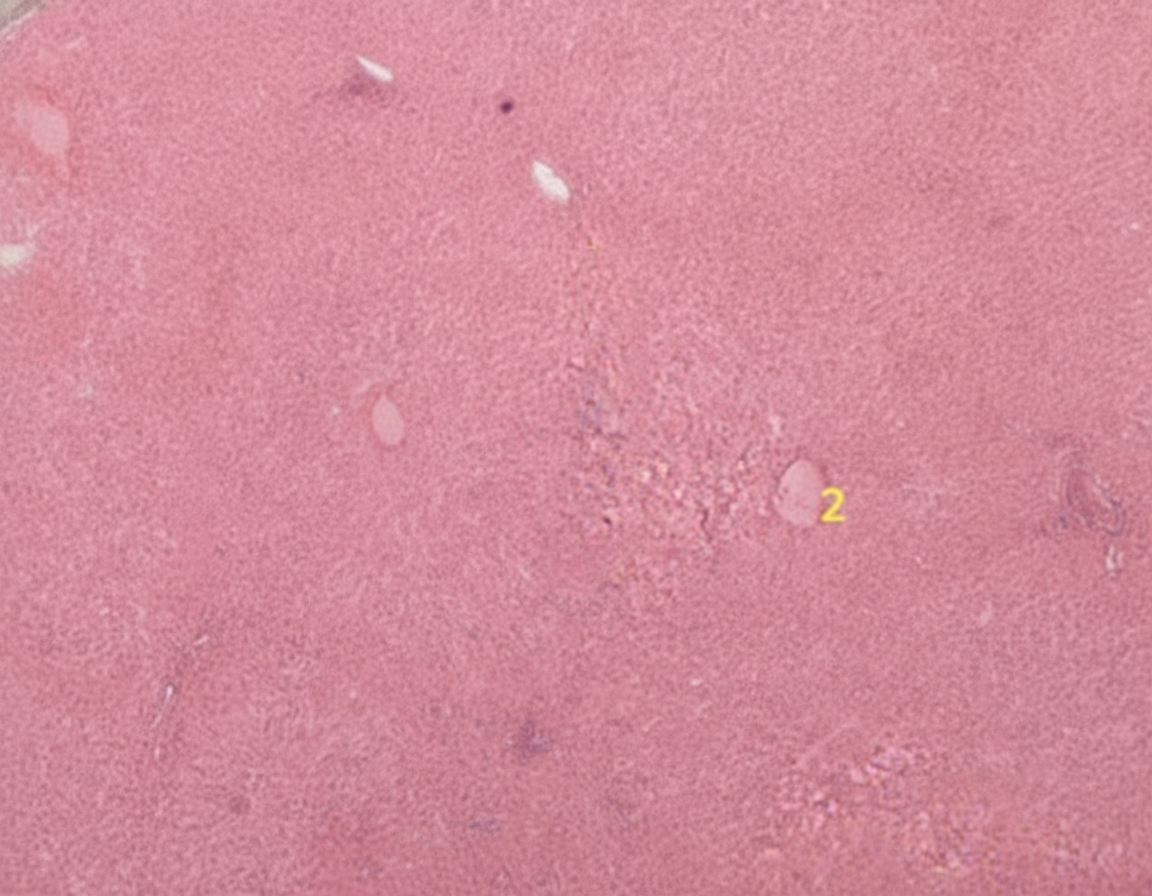
What is the predominant source of blood supply to the organ shown in the histological slide?

What is the hardest and most highly mineralized substance of a tooth?
Corpora amylacea are microscopic structures typically found in which of the following tissues?
Which of the following bone cells has the MOST abundant rough endoplasmic reticulum, Golgi apparatus, and secretory granules?
What type of collagen is primarily found at the dermo-epidermal junction?
Which of the following characteristics is NOT common to both smooth and cardiac muscle?
After maturation in the thymus and release into the circulation, T lymphocytes preferentially migrate to which of the following sites?
Which among the following is the area of greatest demineralization in enamel caries?
What is the epithelial lining of the vagina?
Explanation: ### Explanation The correct answer is **Vagina**. While this may seem counterintuitive because the vaginal lining is composed of **non-keratinized stratified squamous epithelium** (which lacks intrinsic glands), the question asks which structure *classically displays* mucin-producing glands in a clinical/histological context. 1. **Why Vagina is Correct:** Under normal physiological conditions, the vagina does not contain glands [1]. However, the vaginal surface is lubricated by mucus produced by the **cervical glands** [2] and Greater Vestibular (Bartholin) glands [3]. In the context of medical exams, the "mucin-producing" nature of the vaginal environment is a high-yield point often used to differentiate it from other squamous-lined tubes. Furthermore, ectopic mucin-producing glands (Vaginal Adenosis) can occur, particularly in those exposed to DES *in utero*. 2. **Why the others are incorrect:** * **Cervix:** The endocervix contains branched tubular glands that produce mucus [2], but the ectocervix is squamous. While it produces mucus, the vagina is the "classic" answer in specific MCQ formats focusing on the source of vaginal moisture. * **Oesophagus:** Contains esophageal glands (proper and cardiac), but these are primarily seromucous and localized to the submucosa, not the defining feature of the organ's surface. * **Duodenum:** Characterized by **Brunner’s glands** in the submucosa, which produce an alkaline secretion (bicarbonate-rich) to neutralize gastric acid, rather than "mucin" as their primary diagnostic descriptor. ### NEET-PG High-Yield Pearls: * **Vaginal pH:** Normally 3.8–4.5, maintained by **Lactobacillus acidophilus** (Doderlein bacilli) converting glycogen from shed squamous cells into lactic acid. * **Histology Tip:** The vagina lacks a muscularis mucosae, distinguishing it from the esophagus. * **Clinical Correlation:** **Vaginal Adenosis** is the presence of glandular columnar epithelium (mucin-producing) in the vagina, a precursor to Clear Cell Adenocarcinoma.
Explanation: ***Portal vein*** - Supplies **75-80%** of the liver's blood supply, carrying **nutrient-rich deoxygenated blood** from the gastrointestinal tract and spleen. - Forms the **portal triad** along with hepatic artery and bile duct, visible in liver lobule histology as part of the **portal area**. *Hepatic artery* - Provides only **20-25%** of liver blood supply, carrying **oxygenated blood** from the systemic circulation. - While essential for **oxygen delivery** to hepatocytes, it is not the predominant source of blood supply. *Hepatic vein* - Functions as **drainage** rather than supply, carrying deoxygenated blood away from the liver to the **inferior vena cava**. - Located at the **center of liver lobules** (central vein) and represents outflow, not inflow of blood. *Cystic artery* - Supplies blood specifically to the **gallbladder**, not the liver parenchyma. - A branch of the **right hepatic artery**, it has no role in hepatic blood supply and would not be visible in liver histology.
Explanation: **Explanation:** **Enamel** is the correct answer because it is the most highly mineralized and hardest tissue in the human body. It is composed of approximately **96% inorganic material** (primarily hydroxyapatite crystals) and only 4% organic matrix and water. It is derived from the **ectoderm** (specifically from ameloblasts), making it unique among dental tissues. **Analysis of Incorrect Options:** * **Dentine:** While harder than bone, it is less mineralized than enamel (approx. 70% inorganic). It forms the bulk of the tooth and is sensitive to pain. It is derived from the mesoderm (odontoblasts). * **Cementum:** This bone-like substance covers the root of the tooth. It is less mineralized than dentine (approx. 45-50% inorganic) and serves to anchor the periodontal ligament. * **Pulp:** This is the only non-mineralized tissue of the tooth. It is a soft connective tissue containing blood vessels, nerves, and lymphatics, occupying the central pulp cavity. **High-Yield Clinical Pearls for NEET-PG:** * **Ameloblasts:** These cells form enamel but are lost after tooth eruption; therefore, enamel cannot regenerate or repair itself. * **Striae of Retzius:** These are incremental growth lines seen in tooth enamel, representing its rhythmic deposition. * **Fluorosis:** Excessive fluoride intake during enamel formation leads to "mottled enamel" due to hypomineralization. * **Hardness Scale:** Enamel (Hardest) > Dentine > Cementum > Bone.
Explanation: **Explanation:** **Corpora amylacea** (also known as amyloid bodies) are small, laminated hyaline masses found in the acini of the **prostate gland**. They are formed by the calcification of proteinaceous secretions and desquamated epithelial cells. These structures are a characteristic histological feature of the aging prostate and are frequently seen in cases of Benign Prostatic Hyperplasia (BPH). **Analysis of Options:** * **A. Prostate (Correct):** Corpora amylacea are most abundant in the prostatic alveoli. With age, they may undergo calcification to form "prostatic calculi." * **B. Seminal vesicle:** While these glands produce the majority of seminal fluid (rich in fructose), they do not contain laminated corpora amylacea. * **C. Thymus:** The characteristic microscopic feature of the thymus is **Hassall’s corpuscles** (concentric layers of degenerating epithelial reticular cells), not corpora amylacea. * **D. Testis:** The testis is characterized by seminiferous tubules and Leydig cells. It does not contain these laminated proteinaceous bodies. **High-Yield Clinical Pearls for NEET-PG:** * **Staining:** Despite the name "amylacea," these bodies are proteinaceous, though they may stain with iodine (similar to starch). * **CNS Connection:** Corpora amylacea are also found in the **Central Nervous System** (specifically in the end-feet of astrocytes) and increase with age or neurodegenerative diseases. * **Histology Identification:** On H&E stain, they appear as eosinophilic, concentric, "tree-ring" like structures within the glandular lumen. * **Prostatic Acid Phosphatase (PAP):** This is a marker for prostatic tissue, often elevated in prostate cancer.
Explanation: The correct answer is **Osteoblasts**. This question tests your understanding of the correlation between cellular ultrastructure and function in bone tissue. **1. Why Osteoblasts are correct:** Osteoblasts are the "bone-forming" cells [1]. Their primary function is the synthesis and secretion of the organic components of the bone matrix (**osteoid**), which consists mainly of Type I collagen and non-collagenous proteins (like osteocalcin) [1]. To perform this high-volume protein synthesis and secretion, the cell requires: * **Abundant Rough Endoplasmic Reticulum (RER):** For the translation and folding of collagen proteins. * **Prominent Golgi Apparatus:** For the packaging and modification of proteins. * **Secretory Granules:** To transport the matrix components to the cell surface for exocytosis. **2. Why the other options are incorrect:** * **Osteocytes (A):** These are mature bone cells trapped in lacunae. They maintain the matrix and act as mechanosensors. Compared to osteoblasts, they have significantly reduced organelles, reflecting lower metabolic activity. * **Osteoprogenitor Cells (B):** These are mesenchymal stem cells [1]. While they have the potential to become osteoblasts, they are relatively quiescent and lack the specialized protein-synthetic machinery seen in active osteoblasts. * **Bone Lining Cells (C):** These are inactive osteoblasts found on the surface of bones where remodeling is not occurring [1]. They are flattened cells with very few organelles. **High-Yield NEET-PG Pearls:** * **Osteoblasts** are derived from **mesenchymal stem cells**, whereas **Osteoclasts** (bone-resorbing cells) are derived from the **monocyte-macrophage lineage** (hematopoietic) [2]. * **Osteoclasts** are characterized by a "ruffled border" and an abundance of **lysosomes** and mitochondria, rather than RER [2]. * **Marker of Osteoblast activity:** Serum Alkaline Phosphatase (ALP) and Osteocalcin [1].
Explanation: The dermo-epidermal junction (DEJ) is a complex basement membrane zone that anchors the epidermis to the underlying dermis. **Why Type VII is correct:** **Type VII collagen** is the primary component of **anchoring fibrils**. These fibrils originate from the lower part of the basement membrane (lamina densa) and extend into the papillary dermis, looping around Type I and III collagen bundles. This structural arrangement provides the mechanical stability required to prevent the epidermis from shearing off the dermis. **Analysis of Incorrect Options:** * **Type IV Collagen:** While found in the DEJ, it is the main structural component of the **lamina densa** (basal lamina) itself, forming a mesh-like scaffold rather than the specific anchoring fibrils. * **Type XV Collagen:** This is a "multiplexin" collagen found in the vascular and epithelial basement membranes, but it does not play a primary role in the mechanical anchoring of the DEJ. * **Type XXII Collagen:** This is a FACIT collagen primarily localized at the **myotendinous junction**, not the dermo-epidermal junction. **High-Yield Clinical Pearls for NEET-PG:** * **Dystrophic Epidermolysis Bullosa (DEB):** This condition is caused by mutations in the **COL7A1 gene**, leading to defective Type VII collagen. Clinically, it presents with skin fragility and subepidermal blistering. * **Bullous Pemphigoid:** The target antigens are BP180 (Type XVII collagen) and BP230. * **Mnemonic for Collagen Types:** * Type **I**: **B**one (Skin, Tendon) * Type **II**: **C**artilage * Type **III**: **R**eticular fibers (Blood vessels) * Type **IV**: **F**loor (Basement membrane) * Type **VII**: **A**nchoring fibrils (DEJ)
Explanation: ### Explanation The correct answer is **Branching fibers**. While both smooth and cardiac muscles share several histological features, their structural architecture differs significantly. **1. Why "Branching fibers" is the correct answer:** Branching is a hallmark histological feature of **cardiac muscle** fibers [2], allowing them to form a complex three-dimensional network for synchronized contraction. In contrast, **smooth muscle** cells are spindle-shaped (fusiform) with tapered ends and do not branch; they are arranged in sheets or bundles [3]. **2. Analysis of Incorrect Options:** * **Gap Junctions:** These are present in both. In cardiac muscle, they are found in the intercalated discs (specifically the *macula adherens* and *gap junctions*). In smooth muscle (specifically the single-unit type), they allow for functional syncytium [1]. * **Central Nuclei:** Both muscle types typically possess a single, centrally located nucleus. This distinguishes them from skeletal muscle, which has multiple peripherally located nuclei. * **Perinuclear Organelles:** In both smooth and cardiac muscles, organelles such as mitochondria, the Golgi apparatus, and glycogen granules are concentrated in the sarcoplasmic region immediately surrounding the nucleus (the perinuclear zone). **Clinical Pearls for NEET-PG:** * **Skeletal Muscle:** Striated, voluntary, peripheral nuclei, no gap junctions. * **Cardiac Muscle:** Striated, involuntary, central nucleus, **branching**, intercalated discs [2]. * **Smooth Muscle:** Non-striated, involuntary, central nucleus, **fusiform shape**, no T-tubules (uses caveolae instead) [3]. * **High-Yield Fact:** Cardiac muscle is the only muscle type that exhibits both striations and branching. If a question mentions "intercalated discs," it is pathognomonic for cardiac muscle.
Explanation: ### Explanation The correct answer is **A. Paracortex of lymph nodes.** **Concept:** Lymphoid organs are organized into specific zones populated by either B or T lymphocytes. After maturing in the thymus, T cells enter the circulation and home to **thymus-dependent zones** of secondary lymphoid organs [1], [2]. In the lymph node, this specific region is the **paracortex** (the zone between the outer cortex and the medulla). This area is characterized by the presence of **High Endothelial Venules (HEVs)**, which express specific adhesion molecules (addressins) that allow circulating T cells to extravasate into the node [1]. **Analysis of Incorrect Options:** * **B. Cortical lymphoid nodules:** These are located in the outer cortex and are **thymus-independent zones**. They are primarily composed of B lymphocytes. Primary follicles contain resting B cells, while secondary follicles contain germinal centers for B-cell proliferation. * **C. Hilum of lymph nodes:** This is the structural exit point where efferent lymphatic vessels and blood vessels leave/enter the node. It is not a site of lymphocyte sequestration or functional organization. * **D. Lymphoid nodules of the tonsils:** Similar to the nodules in lymph nodes, these are B-cell rich areas. While tonsils do have T-cell zones (interfollicular regions), the paracortex of the lymph node is the classic, high-yield example of a T-cell dependent zone. **NEET-PG High-Yield Pearls:** 1. **Thymus-Dependent Zones:** Paracortex (Lymph Node), Periarteriolar Lymphoid Sheaths (PALS) in the Spleen, and interfollicular areas in Peyer’s patches. 2. **DiGeorge Syndrome:** Due to thymic hypoplasia, the paracortex of lymph nodes and PALS of the spleen will be depleted/underdeveloped. 3. **HEVs:** Found in all secondary lymphoid organs except the spleen [1]. They are the primary entry point for lymphocytes from the blood.
Explanation: **Explanation:** Enamel caries is a dynamic process characterized by distinct histological zones when viewed under polarized light. These zones represent varying degrees of mineral loss and pore volume. **Why "Body of the Lesion" is correct:** The **Body of the Lesion** is the largest portion of incipient enamel caries. It represents the area of **maximum demineralization**, with a mineral loss of approximately **25% to 50%**. Histologically, it has a pore volume of 5% to 25%, making it the most porous and structurally compromised part of the lesion. **Analysis of Incorrect Options:** * **Translucent Zone (A):** This is the advancing front of the lesion. It is the first observable change but shows the **least demineralization** (only about 1% mineral loss). * **Dark Zone (B):** Located between the translucent zone and the body of the lesion, it represents an area of **remineralization**. It has a pore volume of 2% to 4%. * **Surface Zone (D):** This is the relatively intact outer layer of enamel (approx. 30–100 μm thick). Despite the underlying decay, it remains highly mineralized (less than 10% loss) due to constant exposure to fluoride and minerals in saliva. **NEET-PG High-Yield Facts:** * **Order of zones (from deep to superficial):** Translucent zone → Dark zone → Body of the lesion → Surface zone. * **Pore Volume sequence:** Translucent (1%) < Dark (2-4%) < Surface (1-10%) < **Body (5-25%)**. * The **Dark Zone** is "dark" because its tiny pores are filled with air or vapor, which does not transmit light well. Its presence indicates a slower-advancing or arresting lesion.
Explanation: The vaginal lining is a classic high-yield topic in NEET-PG anatomy and histology. Here is the breakdown: ### **Why Option A is Correct** The vagina is lined by **stratified squamous non-keratinized epithelium** [2]. This multi-layered structure is designed to withstand **frictional stress** during intercourse and childbirth. Unlike the skin, it remains moist due to secretions from the cervix and vestibular glands (the vaginal mucosa itself lacks glands) [3]. Under the influence of **estrogen**, these epithelial cells accumulate **glycogen**. When these cells desquamate, the commensal *Lactobacillus acidophilus* (Döderlein’s bacilli) ferments the glycogen into lactic acid, maintaining a protective acidic pH (3.8–4.5) that inhibits pathogen growth. ### **Why Other Options are Incorrect** * **B. Stratified squamous keratinised:** This is found in the **skin (epidermis)** [2]. Keratin provides a waterproof, protective barrier against desiccation, which is unnecessary for the internal moist environment of the vagina. * **C. Columnar:** Simple columnar epithelium lines the **endocervix, uterus, and fallopian tubes** [1]. The transition from columnar (endocervix) to stratified squamous (ectocervix/vagina) occurs at the **Squamocolumnar Junction**, a critical site for cervical cancer screening. * **D. Cuboidal:** Simple cuboidal epithelium is typically found in **glandular ducts** [3] or the **germinal epithelium of the ovary**, not in areas requiring protection against friction. ### **High-Yield Clinical Pearls for NEET-PG** 1. **Embryology:** The upper 1/3rd of the vagina is derived from **Müllerian ducts**, while the lower 2/3rd is derived from the **Urogenital sinus** [2]. 2. **Vaginal Smear:** During the ovulatory phase, high estrogen causes "cornification" (pyknotic nuclei) of these squamous cells. 3. **Transformation Zone:** The area where the epithelium changes from columnar to squamous is the most common site for **Cervical Intraepithelial Neoplasia (CIN)**.
Cellular Ultrastructure
Practice Questions
Microscopic Anatomy of Epithelial Tissues
Practice Questions
Microscopic Anatomy of Connective Tissues
Practice Questions
Microscopic Anatomy of Muscle Tissues
Practice Questions
Microscopic Anatomy of Nervous Tissues
Practice Questions
Microscopic Anatomy of Blood and Immune System
Practice Questions
Microscopic Anatomy of Endocrine Glands
Practice Questions
Microscopic Anatomy of Digestive System
Practice Questions
Microscopic Anatomy of Respiratory System
Practice Questions
Microscopic Anatomy of Urinary System
Practice Questions
Microscopic Anatomy of Reproductive System
Practice Questions
Techniques in Microscopic Anatomy
Practice Questions
Get full access to all questions, explanations, and performance tracking.
Start For Free