
Microscopic Anatomy — MCQs

On this page
Compound microscope was significantly improved and popularized by:
Dense irregular connective tissue is found in:
Langerhans cells are seen in which layer of skin?
Schwalbe's ring corresponds to:
Goblet cells are seen in -
What is the lining of the lacrimal gland alveoli?
Which of the following layers is absent in the esophagus?
Hering's canals are present in ?
In which layer of the gastrointestinal tract is Auerbach's plexus located?
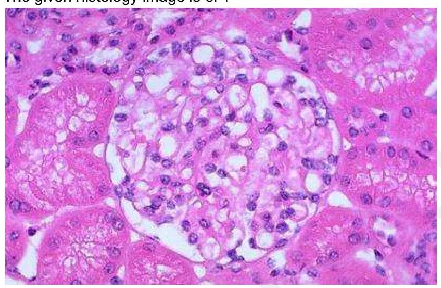
The given histology image is of which structure?
Practice by Chapter
Cellular Ultrastructure
Practice Questions
Microscopic Anatomy of Epithelial Tissues
Practice Questions
Microscopic Anatomy of Connective Tissues
Practice Questions
Microscopic Anatomy of Muscle Tissues
Practice Questions
Microscopic Anatomy of Nervous Tissues
Practice Questions
Microscopic Anatomy of Blood and Immune System
Practice Questions
Microscopic Anatomy of Endocrine Glands
Practice Questions
Microscopic Anatomy of Digestive System
Practice Questions
Microscopic Anatomy of Respiratory System
Practice Questions
Microscopic Anatomy of Urinary System
Practice Questions
Microscopic Anatomy of Reproductive System
Practice Questions
Techniques in Microscopic Anatomy
Practice Questions
Want unlimited practice?
Get full access to all questions, explanations, and performance tracking.
Scan to download app