
Microscopic Anatomy — MCQs

On this page
Which type of collagen forms the basement membrane of the kidney?
The macula densa is derived from which part of the nephron?
Cervical stroma consists of which type of glands?
What type of epithelium lines the maxillary sinus?
The epithelial lining of the cervical canal is:
Serous demilunes are present in large numbers in which gland?
Which banding technique is most commonly employed for cytogenetic analysis?
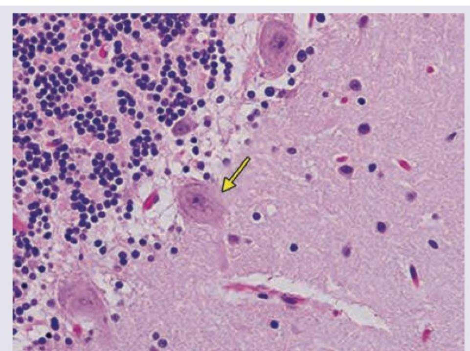
In the given slide of cerebellum, the marked cell is inhibitory to?
The stereocilia of hair cells are embedded in which membrane?
The organ of Corti normally contains:
Practice by Chapter
Cellular Ultrastructure
Practice Questions
Microscopic Anatomy of Epithelial Tissues
Practice Questions
Microscopic Anatomy of Connective Tissues
Practice Questions
Microscopic Anatomy of Muscle Tissues
Practice Questions
Microscopic Anatomy of Nervous Tissues
Practice Questions
Microscopic Anatomy of Blood and Immune System
Practice Questions
Microscopic Anatomy of Endocrine Glands
Practice Questions
Microscopic Anatomy of Digestive System
Practice Questions
Microscopic Anatomy of Respiratory System
Practice Questions
Microscopic Anatomy of Urinary System
Practice Questions
Microscopic Anatomy of Reproductive System
Practice Questions
Techniques in Microscopic Anatomy
Practice Questions
Want unlimited practice?
Get full access to all questions, explanations, and performance tracking.
Scan to download app