All of the following are true about skeletal muscle except?
Which of the following is a layer between choroid and retina?
The organ of Corti normally contains:
Compound microscope was significantly improved and popularized by:
Space of Disse is seen in
Desmosomes are helpful in connecting -
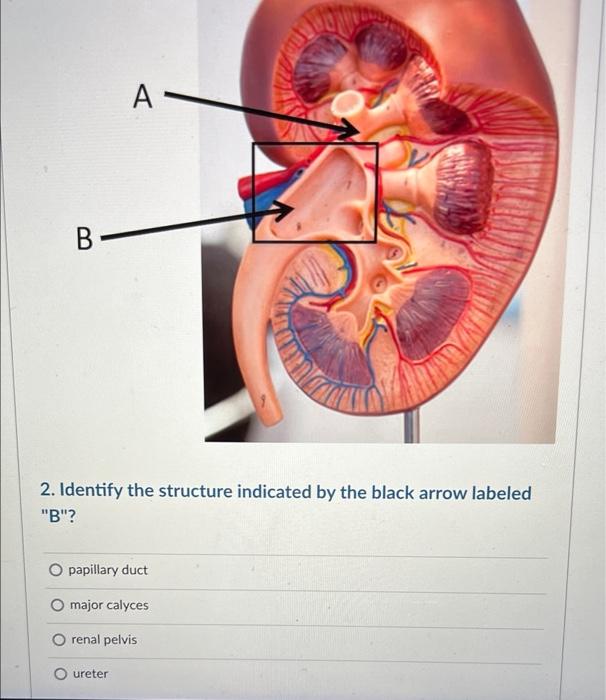
Name the structure marked with an arrow
Dense irregular connective tissue is found in:
Langerhans cells are seen in which layer of skin?
Goblet cells are seen in -
Explanation: The nuclei are central - Skeletal muscle fibers are **multinucleated**, but their nuclei are typically located **peripherally** (just beneath the sarcolemma), not centrally. - This peripheral placement allows the contractile proteins to occupy the majority of the cell's volume. *Have multiple nuclei* - Skeletal muscle cells develop from the fusion of multiple myoblasts, resulting in **syncytia** with numerous nuclei. - This high number of nuclei supports the large volume and metabolic demands of these long, contractile cells. *Have transverse striations* - The organized arrangement of **actin and myosin filaments** into sarcomeres creates a characteristic pattern of light (I bands) and dark (A bands) bands [2]. - These regularly repeating bands are visible as **transverse striations** under a light microscope [1]. *They are cylindrical in shape* - Individual skeletal muscle fibers are typically long and **cylindrical**, maintaining a relatively uniform diameter along their length. - This shape is well-suited for their primary function of generating force and shortening.
Explanation: ***Bruch's membrane*** - **Bruch's membrane** is an acellular barrier located between the **retinal pigment epithelium (RPE)** of the retina and the **choriocapillaris** layer of the choroid [1]. - Its primary function is to serve as a **filtration barrier** and to support the outer retina, playing a crucial role in nutrient and waste exchange [4]. *Photoreceptors* - The **photoreceptor layer** is part of the **retina** itself, containing the rods and cones responsible for light detection, and is not a separate layer between the choroid and retina [2]. - This layer sits **distal to the RPE** and Bruch's membrane, meaning it is closer to the center of the eye. *Descemet's membrane* - **Descemet's membrane** is an elastic and strong basement membrane found in the **cornea**, specifically between the corneal stroma and the corneal endothelium [2]. - It is located at the **front of the eye** and has no relation to the structures between the choroid and retina. *Ganglion cell layer* - The **ganglion cell layer** is a part of the **retina** that contains the cell bodies of the retinal ganglion cells, which transmit visual information from the eye to the brain [3]. - This layer is located **anterior to the photoreceptor layer** and is not situated between the choroid and retina.
Explanation: ***A single row of inner hair cells and 3-4 rows of outer hair cells*** - The **organ of Corti**, crucial for hearing, is structurally organized with a distinct arrangement of hair cells to efficiently transduce sound vibrations into electrical signals. - This specific arrangement ensures optimal **sound processing**, with inner hair cells primarily responsible for converting sound and outer hair cells for amplifying and fine-tuning hearing [1]. *A single row of outer hair cells and 3-4 rows of inner hair cells* - This statement reverses the actual arrangement of hair cells in the cochlea, which is essential for correct auditory function. - The **inner hair cells** are primarily responsible for sound transduction and are less numerous than the **outer hair cells**, which are involved in auditory amplification [1]. *A single row of inner hair cells and a single row of outer hair cells* - While there is indeed a single row of **inner hair cells**, the **outer hair cells** are consistently found in multiple rows (typically three to four) [1]. - This misconception underestimates the complexity and amplification role of the outer hair cells in the auditory system. *3-4 rows each of inner and outer hair cells* - This statement is incorrect because the **inner hair cells** are characteristically arranged in a **single row** [1]. - Having multiple rows of inner hair cells would alter their primary function in **sound transduction** and signal generation for the brain.
Explanation: ***Robert Hooke*** - Robert Hooke significantly improved the compound microscope through his modifications, including using separate lenses for the **objective** and **eyepiece**, and introducing a **lighting system**. - His detailed illustrations and observations, published in *Micrographia*, popularized its use and demonstrated its potential in scientific research, notably his observation of **"cells"** in cork. *Leeuwenhoek* - **Antonie van Leeuwenhoek** is known for his skill in grinding **simple lenses**, designing single-lens microscopes that achieved remarkably high magnifications. - While his microscopes were powerful, they were **single-lens** and not compound microscopes; he is credited with discovering protozoa and bacteria ("animalcules"). *Louis Pasteur* - **Louis Pasteur** was a renowned **chemist and microbiologist** famous for his discoveries related to **vaccinations, microbial fermentation, and pasteurization**. - His work was instrumental in disproving **spontaneous generation** and establishing the germ theory of disease, but he did not significantly improve or popularize the compound microscope itself. *Hippocrates* - **Hippocrates** is an ancient Greek physician, often considered the **"Father of Medicine,"** who lived in the 5th century BC. - His contributions were fundamental to medical ethics and practice, but he predates the invention of the microscope by over two millennia and had no involvement with its development.
Explanation: ***Liver*** - The **Space of Disse**, also known as the perisinusoidal space, is a crucial area located between the **endothelial cells** of the liver sinusoids and the **hepatocytes** [1]. - This space is vital for the exchange of plasma constituents between blood and hepatocytes, containing **hepatic stellate cells (Ito cells)** that store vitamin A and can become fibrogenic in liver injury [1]. *Spleen* - The spleen is characterized by **red pulp** (involved in filtering blood) and **white pulp** (involved in immune responses), without a structure analogous to the Space of Disse. - It contains **splenic cords (cords of Billroth)** and **splenic sinusoids**, which are structurally distinct from the liver's perisinusoidal space. *Kidney* - The kidney's filtering units are **nephrons**, comprising **glomeruli** and **renal tubules**, neither of which feature a Space of Disse. - The kidney has specialized structures like the **Bowman's capsule** and the **juxtaglomerular apparatus** for filtration and regulation, which serve entirely different functions. *Small intestine* - The small intestine is lined with **villi** and **crypts of Lieberkühn**, which are epithelial structures designed for nutrient absorption. - It contains specialized cells like **enterocytes**, **goblet cells**, and **Paneth cells**, but lacks the unique perisinusoidal space found in the liver.
Explanation: ***Keratinocytes*** - **Desmosomes** are specialized cell junctions that provide strong adhesion between adjacent cells, particularly abundant in tissues subject to mechanical stress like the **epidermis** [1]. - They link the **intermediate filaments** (keratin in keratinocytes) of neighboring cells, forming a robust network that resists pulling forces. *Langerhans cells* - **Langerhans cells** are antigen-presenting cells found in the epidermis, but they do not form extensive desmosomal connections with keratinocytes or other cells [1]. - They are primarily involved in immune surveillance and can migrate within the epidermis [1]. *Dermis and epidermis* - The connection between the **dermis and epidermis** is primarily mediated by the **basement membrane**, involving structures like **hemidesmosomes** and anchoring fibrils, not desmosomes between cells. - Hemidesmosomes connect basal keratinocytes to the basement membrane, while desmosomes connect keratinocytes to each other. *Melanocytes* - **Melanocytes** are pigment-producing cells located in the basal layer of the epidermis [1]. - While they are in close proximity to keratinocytes, they do not form significant desmosomal junctions with them; they primarily interact through dendrites that transfer melanin [1].
Explanation: ***Secretory vesicles*** - The image shows an **electron micrograph of a cell**, likely an endocrine or exocrine cell, with numerous small, membrane-bound structures near the plasma membrane. - These structures, indicated by the arrow, are consistent with secretory vesicles, which store and transport substances (e.g., hormones, enzymes) to be released from the cell via **exocytosis**. *Mitochondria* - Mitochondria are **larger organelles** with characteristic inner folds called cristae, and are primarily involved in ATP production. - The structures pointed to by the arrow are much smaller and lack the internal structure typical of mitochondria. *Rough Endoplasmic reticulum* - The rough ER is a network of interconnected membranes studded with ribosomes, involved in protein synthesis and modification. - It usually appears as **flattened sacs or cisternae** and is distinct from the individual, small, spherical structures shown. *Golgi bodies* - The Golgi apparatus consists of **flattened membrane-bound sacs** called cisternae, arranged in stacks, and is involved in processing and packaging proteins and lipids. - While it processes substances destined for secretion, the structures indicated by the arrow are the final transport vesicles, not the Golgi stack itself.
Explanation: ***Dermis*** - The **dermis** is primarily composed of **dense irregular connective tissue**, which provides structural strength and elasticity to the skin. [1] - This tissue consists of a rich network of **collagen fibers** arranged in various directions, along with elastic fibers, offering resistance to stress from multiple directions. [1] *Ligament* - Ligaments are examples of **dense regular connective tissue**, not irregular. - Their collagen fibers are densely packed and arranged in a **parallel fashion** to resist unidirectional stress. *Lamina propria* - The lamina propria is a layer of **loose connective tissue** underlying the epithelium of many organs. - It contains a higher proportion of **ground substance** and fewer, more loosely arranged fibers compared to dense connective tissue. *Tendon* - Tendons are also examples of **dense regular connective tissue**, connecting muscle to bone. - They are characterized by **parallel arrays of collagen fibers** adapted to withstand strong tensile forces in a single direction.
Explanation: ***Stratum spinosum*** - **Langerhans cells** are antigen-presenting cells found predominantly in the **stratum spinosum** of the epidermis [1]. - Their dendritic morphology allows them to capture antigens effectively and migrate to lymph nodes to initiate immune responses [1]. *Stratum basale* - This layer primarily contains **basal keratinocytes** responsible for cell division and regeneration of the epidermis, and **melanocytes** which produce melanin [1]. - While Langerhans cells can occasionally be observed in the basal layer, their primary and most abundant location is the stratum spinosum [1]. *Stratum corneum* - The stratum corneum is the **outermost layer** of the epidermis, consisting of flattened, dead keratinocytes filled with keratin [1]. - This layer provides a protective barrier and is devoid of living cells like immune cells. *Stratum granulosum* - This layer is characterized by cells containing **keratohyalin granules** and lamellar bodies, essential for skin barrier function. - While immune cells can traverse different layers, the stratum granulosum is not the primary residence for Langerhans cells.
Explanation: ***Conjunctiva*** - **Goblet cells** are specialized **mucus-secreting cells** found in the conjunctiva, specifically in the **fornices** and along the **palpebral conjunctiva** [1]. - They produce **mucin**, an essential component of the **tear film** that helps in lubrication and protection of the ocular surface [2]. *Cornea* - The cornea is composed primarily of stratified squamous epithelium, Bowman's layer, stroma, Descemet's membrane, and endothelium [1]. - It does **not contain goblet cells**; its primary role is light transmission and refraction [1]. *Retina* - The retina is a **neurosensory tissue** responsible for converting light into neural signals [3]. - It contains various photoreceptor cells (rods and cones) and neurons, but **no goblet cells** [3]. *Vitreous* - The vitreous humor is a **gel-like substance** that fills the space between the lens and the retina. - It is composed mainly of water, collagen, and hyaluronic acid, and **does not contain any cells**, including goblet cells.
Cellular Ultrastructure
Practice Questions
Microscopic Anatomy of Epithelial Tissues
Practice Questions
Microscopic Anatomy of Connective Tissues
Practice Questions
Microscopic Anatomy of Muscle Tissues
Practice Questions
Microscopic Anatomy of Nervous Tissues
Practice Questions
Microscopic Anatomy of Blood and Immune System
Practice Questions
Microscopic Anatomy of Endocrine Glands
Practice Questions
Microscopic Anatomy of Digestive System
Practice Questions
Microscopic Anatomy of Respiratory System
Practice Questions
Microscopic Anatomy of Urinary System
Practice Questions
Microscopic Anatomy of Reproductive System
Practice Questions
Techniques in Microscopic Anatomy
Practice Questions
Get full access to all questions, explanations, and performance tracking.
Start For Free