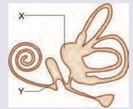
All are correct about the part marked as $X$ and $Y$ except:

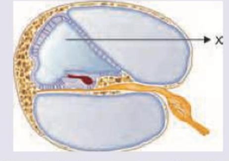
What is correct about the composition of fluid in the area marked as $X$ ?

Cords of Billroth in spleen are found in
Arrange coverings on peripheral nerve from inner to outer
Elastic cartilage is present in
Periosteal matrix acts on bone tissue to bring about:
In which of the following structures is regularly arranged parallel collagen present?
Submucosal plexus is -
The Eustachian tube is made up of
The stereocilia of hair cells are embedded in which membrane?
Explanation: This is an EXCEPT question - we need to identify the **incorrect** statement. ***$X$ is utricle and has anterior upward slope of 30 degrees*** ✓ **INCORRECT STATEMENT - This is the answer** - The **utricle's macula** is oriented **horizontally** when the head is in normal anatomical position, NOT at a 30-degree anterior upward slope - It is the **saccule's macula** that has an orientation closer to vertical (approximately 30° from vertical in some references) - The utricle detects **linear acceleration in the horizontal plane** - **This statement is FALSE, making it the correct answer to this EXCEPT question** *$X$ is utricle and receives 5 openings of three semicircular canals* ✓ **CORRECT STATEMENT** - This is anatomically **correct**. The utricle receives **five openings** from the three semicircular canals - The three semicircular canals (anterior, posterior, and lateral) have five openings because the **anterior and posterior canals share a common crus** - **This statement is TRUE, so it is not the answer** *$Y$ is ductus reuniens* ✓ **INCORRECT STATEMENT (but see note)** - $Y$ appears to point to the **saccule**, which is one of the **otolith organs** in the vestibule - The **ductus reuniens** is a small duct that connects the saccule to the cochlear duct, not the saccule itself - However, **without seeing the image**, if Y points to the ductus reuniens itself, this could be correct - Based on typical anatomy diagrams, Y most likely indicates the saccule, making this statement incorrect *$Y$ connects utricle to cochlear duct* ✓ **INCORRECT STATEMENT** - $Y$ is the **saccule**, not a connecting duct - The saccule connects to the cochlear duct via the **ductus reuniens** - The saccule does NOT directly connect the utricle to the cochlear duct - Anatomically, the utricle and saccule connect via the **utriculosaccular duct** **Key Point**: Since this is an EXCEPT question asking "All are correct EXCEPT", only **Option A** is the definitively FALSE statement about the anatomy. Options B and C are also incorrect statements, but Option A is the clearest incorrect statement based on standard anatomical orientation of the utricle's macula.
Explanation: ***Na = 3 mEq/L, K = 150 mEq/L, Chloride = 125 mEq/L*** - The area marked 'X' represents the **scala media** in the cochlear cross-section, which contains **endolymph**. - Endolymph has a unique ionic composition with **high potassium concentration** (150 mEq/L) and **low sodium concentration** (3 mEq/L), similar to intracellular fluid. *Na = 150 mEq/L, K = 3 mEq/L, Chloride = 125 mEq/L* - This composition represents **perilymph**, found in the **scala vestibuli** and **scala tympani**, not in the scala media. - Perilymph has high Na+ and low K+ concentrations, resembling **cerebrospinal fluid** and extracellular fluid. *Na = 150 mEq/L, K = 30 mEq/L, Chloride = 125 mEq/L* - The **potassium level of 30 mEq/L** is intermediate and doesn't match any physiological cochlear fluid composition. - This value is too high for perilymph (normally 3 mEq/L) and too low for endolymph (normally 150 mEq/L). *Na = 50 mEq/L, K = 30 mEq/L, Chloride = 125 mEq/L* - The **sodium concentration of 50 mEq/L** is abnormally low for any extracellular fluid compartment. - Neither the Na+ nor K+ values correspond to the known ionic gradients essential for **cochlear hair cell function**.
Explanation: ***Correct: Red pulp*** - The **cords of Billroth** (or splenic cords) are a distinctive histological feature of the **red pulp** of the spleen. - These cords consist of a meshwork of reticular cells and reticular fibers, packed with macrophages, lymphocytes, plasma cells, and abundant red blood cells. *Incorrect: White pulp* - The **white pulp** is primarily involved in immune functions, containing lymphoid follicles rich in lymphocytes. - It is organized around central arterioles and does not contain the cords of Billroth. *Incorrect: Trabecular zone* - The **trabecular zone** consists of connective tissue septa that extend inward from the splenic capsule, carrying blood vessels and nerves. - It provides structural support to the spleen but is not where the cords of Billroth are located. *Incorrect: Mantle zone* - The **mantle zone** is a region within the **white pulp**, surrounding the germinal centers of lymphoid follicles. - It is composed mainly of naive B lymphocytes and is distinct from the cords of Billroth.
Explanation: The endoneurium is the innermost delicate connective tissue sheath that surrounds individual nerve fibers (axons), providing structural support and maintaining the microenvironment [1]. The perineurium is a stronger, protective sheath that encircles bundles of nerve fibers, called fascicles, forming a crucial barrier [2]. The epineurium is the outermost, toughest connective tissue layer that surrounds the entire peripheral nerve, encompassing multiple fascicles and their surrounding perineurium.
Explanation: ***Epiglottis*** - The **epiglottis** is composed of **elastic cartilage**, which provides flexibility to fold over the glottis during swallowing, preventing food from entering the trachea. - This type of cartilage is characterized by a dense network of **elastic fibers** within its matrix, allowing it to spring back to its original shape. *Cricoid* - The **cricoid cartilage** is a ring-shaped cartilage that forms the inferior wall of the larynx and is composed of **hyaline cartilage**. - **Hyaline cartilage** provides structural support and maintains the patency of the airway, but lacks the flexibility of elastic cartilage. *Arytenoid cartilage* - The **arytenoid cartilages** are small, paired cartilages that sit atop the cricoid cartilage and are primarily composed of **hyaline cartilage**. - They play a crucial role in the movement of the **vocal cords**, but their limited flexibility is not characteristic of elastic cartilage. *Thyroid cartilage* - The **thyroid cartilage**, the largest laryngeal cartilage, forms the "Adam's apple" and is made of **hyaline cartilage**. - Its rigid structure provides protection for the vocal cords and maintains the shape of the larynx, which is a function of hyaline rather than elastic cartilage.
Explanation: ***Bone modeling*** - The **periosteum** is a vital outer layer of bone that plays a crucial role in bone growth, repair, and **modeling** [1]. - **Bone modeling** involves changes in bone size and shape, often through independent processes of bone formation and resorption on different surfaces, which the periosteal matrix actively contributes to [1]. *Bone resorption only* - While **bone resorption** is a component of bone remodeling, the periosteal matrix is involved in both bone formation and resorption, not exclusively resorption [2]. - Focusing solely on resorption neglects the periosteum's role in **bone deposition** and overall bone shape changes [1]. *Cartilage formation* - The periosteum does not primarily lead to **cartilage formation**; its main role is in bone tissue [3]. - Cartilage formation is typically mediated by chondrocytes, usually in different developmental contexts or repair processes [4]. *Bone mineralization only* - **Bone mineralization** is the process by which inorganic minerals (chiefly calcium phosphate) are deposited into the organic matrix of bone [3]. - While the periosteum contributes to the formation of the organic matrix, its role extends beyond just mineralization to the overall **structuring and reshaping of bone** [1].
Explanation: ### Cornea - The cornea contains **Type I collagen fibers** that are arranged in a precise, orthogonal, and parallel pattern within its lamellae [1]. - This highly organized arrangement is crucial for its **transparency** and its ability to refract light effectively [1]. *Basement membrane* - The basement membrane consists primarily of **Type IV collagen**, **laminin**, and **heparan sulfate proteoglycans** [2]. - Its collagen fibers form a **mesh-like network** rather than a regularly arranged parallel structure [2]. *Diaphragm* - The diaphragm is composed of **skeletal muscle tissue**, which contains contractile proteins (actin and myosin) and connective tissue elements like **endomysium**, **perimysium**, and **epimysium**. - While it contains collagen, it is not arranged in the highly parallel, organized fashion found in structures like the cornea; its primary function is muscle contraction, not transparency or tensile strength from parallel fibers. *Tympanic membrane* - The tympanic membrane (eardrum) has a fibrous middle layer that is largely composed of **collagen fibers**. - These fibers are generally arranged in **radial and circular patterns** to provide tensile strength and flexibility for sound transmission, but not in the regular, parallel arrangement characteristic of the cornea.
Explanation: ***Meissner's plexus*** - The **submucosal plexus** is also known as **Meissner's plexus**, located in the submucosal layer of the **gastrointestinal tract** [1]. - It primarily controls local **secretions**, **absorption**, and **blood flow** within the gut [2]. *Myenteric plexus* - The **myenteric plexus** is also known as **Auerbach's plexus**, located between the longitudinal and circular muscle layers of the **gastrointestinal tract** [1]. - It primarily controls **gastrointestinal motility**, not local secretions [1]. *Tympanic plexus* - The **tympanic plexus** is a network of nerves in the **middle ear** that provides sensory innervation to the tympanic cavity. - It is unrelated to the gastrointestinal tract or its intrinsic nervous system. *Auerbach's plexus* - **Auerbach's plexus** is another name for the **myenteric plexus**, which is located between the muscle layers [1]. - It is responsible for gut motility and distinct from the submucosal plexus.
Explanation: ***Elastic cartilage*** - The **cartilaginous portion** of the Eustachian tube (medial 2/3) is primarily composed of **elastic cartilage**. - This tissue provides the necessary **flexibility and resilience** for tube opening during swallowing and yawning, which is essential for **middle ear ventilation**. - Elastic cartilage contains abundant **elastic fibers** that allow the tube to change shape and return to its original position. *Fibro-elastic cartilage* - This term is sometimes used descriptively to refer to **elastic cartilage with fibrous components**, which is what forms the Eustachian tube. - However, as a standalone option separate from "elastic cartilage," it may cause confusion since the standard classification recognizes elastic cartilage as the primary cartilage type. - The cartilaginous Eustachian tube is best classified as **elastic cartilage**. *Hyaline cartilage* - **Hyaline cartilage** is found in the nose, trachea, larynx, and articular surfaces of joints. - It has a smooth, glassy appearance but lacks the **elastic fiber content** needed for the dynamic function of the Eustachian tube. - Would not provide the necessary **flexibility for tube opening** required during pressure equalization. *Fibrocartilage* - **Fibrocartilage** is found in intervertebral discs, pubic symphysis, and menisci, providing **high tensile strength**. - While durable, it lacks the **elastic recoil** necessary for the Eustachian tube to repeatedly open and close. - The Eustachian tube requires **flexibility with elastic recovery**, not primarily tensile strength.
Explanation: ***Tectorial*** - The **stereocilia** of the hair cells of the organ of Corti are directly embedded in the **tectorial membrane** [1]. - This contact is crucial for the transduction of sound vibrations into electrical signals as it allows for the bending of stereocilia when the basilar membrane vibrates [1]. *Reissner's* - **Reissner's membrane** (also known as the vestibular membrane) separates the scala vestibuli from the scala media. - Its primary role is to maintain the difference in ion concentrations between the endolymph and perilymph, and it does not directly interact with hair cell stereocilia. *Tympanic* - The **tympanic membrane** (eardrum) is located at the entrance of the middle ear and vibrates in response to sound waves [1]. - It transmits these vibrations to the ossicles, but it does not contain hair cells or stereocilia [1]. *Basilar* - **Basilar membrane** supports the organ of Corti, which includes the hair cells. - While the hair cells rest on the basilar membrane, their stereocilia are not embedded within it; rather, the basilar membrane's vibration causes the shear force that bends the stereocilia.
Cellular Ultrastructure
Practice Questions
Microscopic Anatomy of Epithelial Tissues
Practice Questions
Microscopic Anatomy of Connective Tissues
Practice Questions
Microscopic Anatomy of Muscle Tissues
Practice Questions
Microscopic Anatomy of Nervous Tissues
Practice Questions
Microscopic Anatomy of Blood and Immune System
Practice Questions
Microscopic Anatomy of Endocrine Glands
Practice Questions
Microscopic Anatomy of Digestive System
Practice Questions
Microscopic Anatomy of Respiratory System
Practice Questions
Microscopic Anatomy of Urinary System
Practice Questions
Microscopic Anatomy of Reproductive System
Practice Questions
Techniques in Microscopic Anatomy
Practice Questions
Get full access to all questions, explanations, and performance tracking.
Start For Free