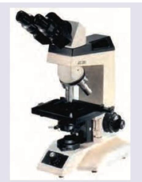
The following image shows:

Identify the test shown in the image provided:

All are true about the anaerobic jar shown below except:

The image shows:

The following instrument is sterilized by:

Which of the following bacteria will have following appearance?

Based on the flagellar arrangement shown in the image, the bacterial flagellation type is:

Which of the following bacteria will have following appearance?

Which of the following is correct about the image shown?

All are true about the instrument shown except:

Explanation: ***Transduction*** - The image illustrates a **bacteriophage** injecting its DNA into a donor bacterial cell, leading to the packaging of bacterial DNA into new phage particles. - Subsequently, these phages infect a recipient cell, transferring the **donor bacterial DNA** and indicating genetic transfer mediated by a virus. *Transformation* - This process involves the direct uptake of **naked extracellular DNA** by competent bacterial cells. - The image clearly depicts a **phage (virus)** mediating the transfer, not naked DNA uptake. *Lysogenic conversion* - This occurs when a bacterium acquires new characteristics due to the presence of a **prophage (integrated bacteriophage DNA)**, which does not necessarily involve general transfer of host DNA to another bacterium. - The image shows the transfer of bacterial DNA from one bacterium to another via a phage, rather than a change in phenotype due to prophage integration. *Conjugation* - This is the direct transfer of genetic material between bacterial cells through cell-to-cell contact, typically via a **pilus**. - The image shows a **phage** as the vector for DNA transfer, not direct bacterial contact.
Explanation: ***Indole test*** - The image displays a reaction where a **red ring** has formed at the surface of the medium in the left tube, while the right tube shows no such color change, appearing yellow. This red ring signifies a **positive indole test**, indicating the presence of **tryptophanase** enzyme. - The Indole test detects an organism's ability to **metabolize tryptophan** into indole, pyruvate, and ammonia using the enzyme **tryptophanase**, with indole being detected by Kovac's reagent forming a red layer. *Methyl red test* - The Methyl Red test detects organisms that produce and maintain **stable acid end-products** from glucose fermentation, resulting in a **red color** if positive or yellow if negative. - The color change in the image is a red ring at the top, not a uniform red color throughout the medium, which suggests a different test. *Nitrate reduction test* - The nitrate reduction test checks for the presence of **nitrate reductase**, an enzyme that converts **nitrate to nitrite** or beyond. - Positive results are typically indicated by a red color after adding reagents A and B, or gas production, which is not depicted in this image. *Voges-Proskauer test* - The Voges-Proskauer (VP) test identifies bacteria that produce **acetoin** from glucose fermentation. - A positive result is indicated by a **red color** developing after adding **Barritt's reagents**, which is different from the distinct red ring formation seen in the image.
Explanation: *Reduced methylene blue is used as indicator* - This statement is incorrect because **methylene blue** is used as an **oxidation-reduction indicator** in anaerobic jars, but in its oxidized (blue) form. It turns colorless when reduced in the absence of oxygen. - The indicator needs to be in its **oxidized state initially** to accurately show the depletion of oxygen; if it were already reduced, it wouldn't indicate proper anaerobic conditions. *McIntosh and Filde's jar* - The image shown is a classic representation of a **McIntosh and Fildes anaerobic jar**, which is widely used for creating an anaerobic environment for culturing anaerobic microorganisms. - This jar is designed with a **catalyst chamber** and a tight-fitting lid to facilitate oxygen removal. *Contains palladinized catalyst for conversion of hydrogen and oxygen to water* - The jar contains a **palladinized catalyst** (e.g., palladium pellets or granules) which plays a crucial role in removing residual oxygen. - This catalyst facilitates the chemical reaction between **added hydrogen** (from a gas pack or direct inlet) and any remaining **oxygen** within the jar to form water, thus creating an anaerobic atmosphere. *Made of glass* - Early anaerobic jars, including the McIntosh and Fildes type, were traditionally made of **thick-walled glass** to withstand the slight vacuum created inside and to allow for visual inspection. - While modern versions may use durable plastics, the classic design, as depicted, is characteristically made of glass.
Explanation: ***Loeffler serum slope*** - This image shows a **Loeffler serum slope**, characterized by its **pale yellow, solidified appearance** in a test tube, often used for culturing *Corynebacterium diphtheriae*. - The medium provides various nutrients and typically encourages the rapid growth of diphtheria bacilli, producing **characteristic morphology**. *Lowenstein Jensen media* - **Lowenstein Jensen media** is typically **greenish** due to the presence of **malachite green**, which inhibits the growth of most contaminants. - It is primarily used for the cultivation of **mycobacteria**, particularly *Mycobacterium tuberculosis*. *Wilson-Blair medium* - **Wilson-Blair medium** is a classic selective medium used for the isolation of *Salmonella typhi* and other *Salmonella* species, appearing as **black colonies**. - This medium contains **bismuth sulfite**, which imparts a distinctive black color to the colonies due to hydrogen sulfide production. *Selenite F broth* - **Selenite F broth** is a **clear enrichment broth** used for the isolation of *Salmonella* and *Shigella* species from fecal samples. - It works by **inhibiting the growth of coliforms** while allowing *Salmonella* and *Shigella* to multiply.
Explanation: ***Ethylene dioxide*** - The image shows a **heart-lung machine (cardiopulmonary bypass machine)**. These complex devices contain various materials, including plastics and electronics, that cannot withstand high temperatures. - **Ethylene oxide (ETO) sterilization** is a low-temperature gaseous sterilization method suitable for heat-sensitive and moisture-sensitive medical devices, making it ideal for the intricate components of a heart-lung machine. *Glutaraldehyde* - **Glutaraldehyde** is a high-level disinfectant and sterilant typically used for instruments that can be **immersed in liquid**, such as endoscopes. - It is not practical or effective for sterilizing large, complex electronic equipment like a heart-lung machine, which has many non-immersible parts. *Aniline dye* - **Aniline dyes** are primarily used as histological stains and in some antiseptic preparations, but they are **not a method of sterilization**. - They lack the broad-spectrum microbicidal activity required for sterilizing medical devices. *Beta-propiolactone* - **Beta-propiolactone** is a chemical sterilant that has been used in the past, primarily for sterilizing vaccines and tissue grafts due to its rapid microbicidal action. - However, its use has been largely **limited due to its carcinogenic properties**, and it is not a common or preferred method for sterilizing large medical equipment like a heart-lung machine.
Explanation: ***Alcaligenes faecalis*** - The diagram shows a bacterium with **lophotrichous flagella**, meaning multiple flagella originate from one or both ends of the cell. This morphology is characteristic of *Alcaligenes faecalis*. - This bacterium is a **gram-negative rod** commonly found in the environment and can be an opportunistic pathogen. *Vibrio cholerae* - *Vibrio cholerae* is characterized by a **single polar flagellum** (monotrichous), giving it a characteristic darting motility. - Its shape is typically **comma-shaped** or curved rod, unlike the straight rod shown with lophotrichous flagella. *Escherichia coli* - *E. coli* exhibits **peritrichous flagella**, meaning flagella are distributed all around the cell surface. - This is different from the lophotrichous arrangement shown in the image where flagella emerge from one or both poles. *Pseudomonas aeruginosa* - *Pseudomonas aeruginosa* typically has **monotrichous flagella** (single polar flagellum) or occasionally **amphitrichous** (one flagellum at each pole). - The image shows multiple flagella at the poles (lophotrichous), which is not characteristic of *Pseudomonas*.
Explanation: ***Peritrichous*** - The image shows numerous flagella **distributed randomly** over the entire surface of the bacterial cell. - This uniform distribution of flagella all around the bacterial body is the defining characteristic of **peritrichous** flagellation. *Monotrichous* - This term describes bacteria that possess a **single flagellum** at one pole of the cell. - The image clearly depicts multiple flagella spread across the cell, not a single polar flagellum. *Lophotrichous* - This refers to bacteria having a **tuft or cluster of flagella** at one or both ends of the cell. - The flagella in the image are distributed across the surface, not concentrated in a bundle at an end. *Amphitrichous* - This describes bacteria with a **single flagellum at each of two opposite ends** of the cell. - The flagella seen in the image are numerous and surround the entire cell, not just at two poles.
Explanation: ***Alcaligenes faecalis*** - The image shown is a **rod-shaped bacterium** with a **tuft of flagella at each pole**, resulting in a **bipolar flagellation** pattern typical of *Alcaligenes faecalis*. - This arrangement of flagella (amphitrichous) is characteristic for motility in this specific bacterium. *Vibrio cholerae* - *Vibrio cholerae* is a **curved rod** (comma-shaped) and typically possesses a **single polar flagellum** (monotrichous), which is different from the structure shown. - Its unique shape and flagellar arrangement are key identifiers in microbiology. *Spirilla* - **Spirilla** are **spiral-shaped bacteria** with a rigid, wavy form, which is distinctly different from the rod shape depicted in the image. - They can have various flagellar arrangements, but their overall morphology is elongated and coiled, not straight. *Salmonella typhi* - *Salmonella typhi* is a **rod-shaped bacterium** but it is typically **peritrichous**, meaning it has flagella distributed all over its cell surface, not just at the poles. - The image clearly shows flagella only at the ends, ruling out *Salmonella typhi*.
Explanation: ***A = Monotrichous, B = Lophotrichous, C = Amphitrichous, D = Peritrichous*** - Image **A** shows a bacterium with a **single flagellum at one pole**, which is the classic presentation of **Monotrichous** flagellation. - Image **B** depicts a bacterium with a **tuft of multiple flagella originating from one pole**, characteristic of **Lophotrichous** arrangement. - Image **C** shows a bacterium with **single flagella at both opposite poles**, consistent with **Amphitrichous** flagellation. - Image **D** demonstrates **flagella distributed uniformly over the entire bacterial cell surface**, which is the defining feature of **Peritrichous** flagellation. - This option correctly identifies all four flagellar arrangements as per standard microbiological classification. *A = Lophotrichous, B = Monotrichous, C = Amphitrichous, D = Peritrichous* - Incorrectly identifies image A as **Lophotrichous** (which requires multiple flagella at one pole), when it clearly shows only a single flagellum. - Also incorrectly labels image B as **Monotrichous** (single flagellum), when it displays a tuft of multiple flagella at one pole. *A = Monotrichous, B = Lophotrichous, C = Peritrichous, D = Amphitrichous* - Incorrectly identifies image C as **Peritrichous**, but the image shows flagella only at two opposite poles, not distributed all over the cell. - Misidentifies image D as **Amphitrichous** (flagella at two poles only), when it clearly shows flagella covering the entire bacterial surface. *A = Peritrichous, B = Lophotrichous, C = Amphitrichous, D = Monotrichous* - Incorrectly labels image A as **Peritrichous** (flagella all over), when it shows a single polar flagellum characteristic of Monotrichous arrangement. - Misidentifies image D as **Monotrichous** (single flagellum), when it demonstrates the peritrichous pattern with flagella distributed over the entire surface.
Explanation: ***Correct Answer: Cannot be used to examine motility of bacteria*** - This statement is **incorrect** (making it the correct answer for this "EXCEPT" question) because the instrument shown is a **light microscope**, which is routinely used to observe the **motility of bacteria** in a wet mount or hanging drop preparation. - Observing bacterial movement, such as **true motility** (swimming) versus **Brownian motion** (random jiggling), is a fundamental microbiological technique facilitated by light microscopes. *Incorrect: Can be used for dark ground microscopic examination of spirochetes* - The instrument is a **light microscope**, and with a specialized **dark-field condenser**, it can be adapted for **dark-field microscopy**. - **Dark-field microscopy** is particularly useful for visualizing unstained, unpigmented, or highly refractile organisms like **spirochetes** (e.g., *Treponema pallidum*), which appear brightly illuminated against a dark background. *Incorrect: Cannot study bacteria less than 0.2 μm* - Light microscopes, including the one pictured, have a **resolution limit** primarily determined by the wavelength of light and the numerical aperture of the objective lens. - The practical resolution limit of a light microscope is generally around **0.2 μm**, meaning objects smaller than this cannot be clearly resolved and appear as blurry dots rather than distinct structures. *Incorrect: Total magnification at 100X objective lens (oil immersion) is 1000 times* - **Total magnification** is calculated by multiplying the magnification of the **objective lens** by the magnification of the **eyepiece** (ocular lens). - Typically, standard microscopes have 10X eyepieces. Therefore, with a **100X oil immersion objective**, the total magnification is 10X (eyepiece) × 100X (objective) = **1000X**.
Cellular Ultrastructure
Practice Questions
Microscopic Anatomy of Epithelial Tissues
Practice Questions
Microscopic Anatomy of Connective Tissues
Practice Questions
Microscopic Anatomy of Muscle Tissues
Practice Questions
Microscopic Anatomy of Nervous Tissues
Practice Questions
Microscopic Anatomy of Blood and Immune System
Practice Questions
Microscopic Anatomy of Endocrine Glands
Practice Questions
Microscopic Anatomy of Digestive System
Practice Questions
Microscopic Anatomy of Respiratory System
Practice Questions
Microscopic Anatomy of Urinary System
Practice Questions
Microscopic Anatomy of Reproductive System
Practice Questions
Techniques in Microscopic Anatomy
Practice Questions
Get full access to all questions, explanations, and performance tracking.
Start For Free