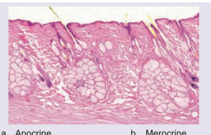
Which type of gland is depicted here?

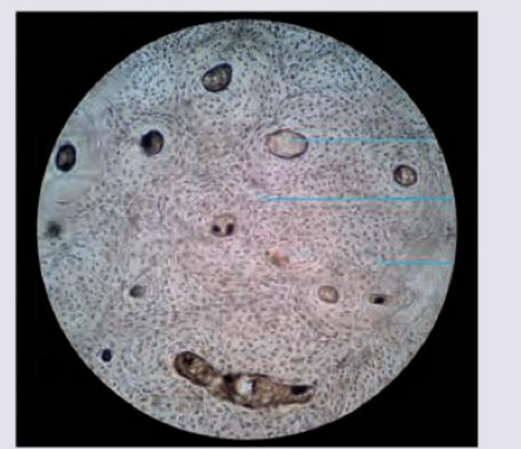
The image shows presence of:

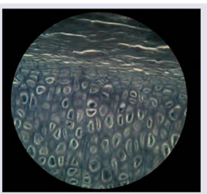
Identify the type of cartilage shown in the figure:

Which of the following has holocrine secretion?
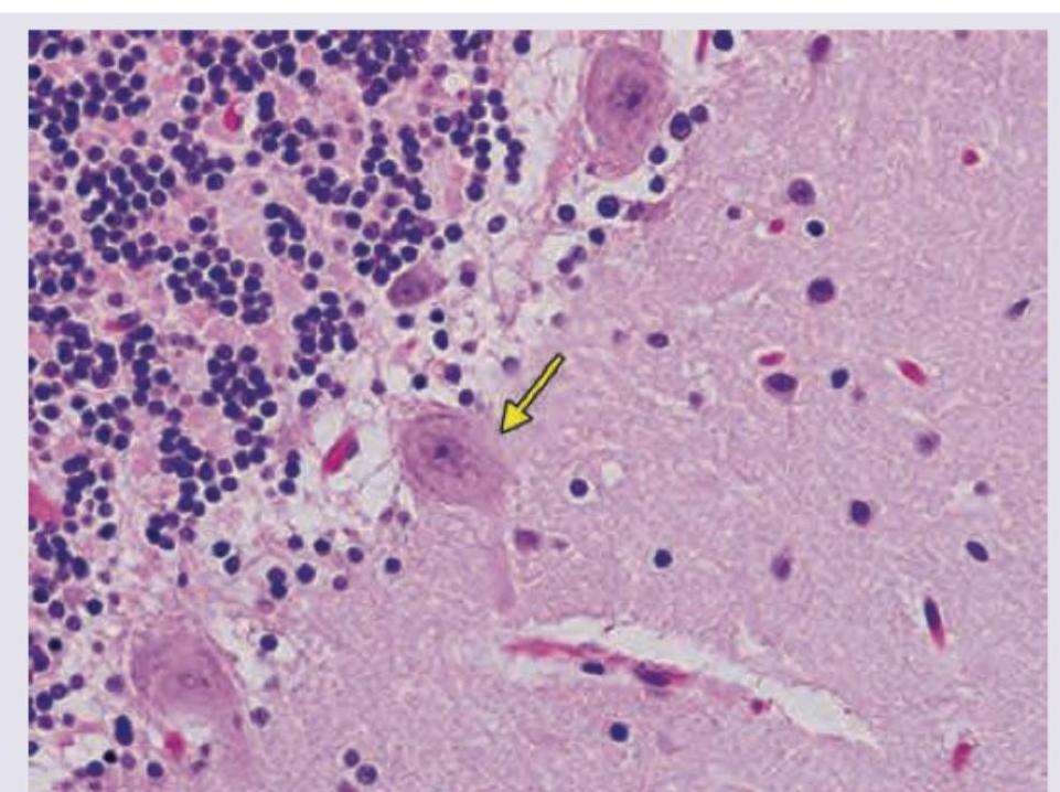
In the given slide of cerebellum, the marked cell is inhibitory to?

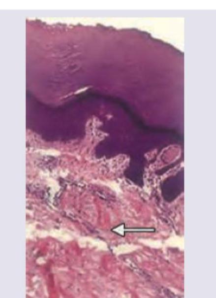
Identify the type of connective tissue present in the area marked with the arrow.

The image shows:

Which is not correct about the following?

The image given below shows:

The following image shows:

Explanation: ***Holocrine*** - The image depicts sebaceous glands, which are **holocrine glands**. In holocrine secretion, the entire cell disintegrates to release its contents. - The cells appear filled with lipid droplets, and as they mature, they move towards the center of the acinus, eventually lysing to release the **sebum** into the hair follicle. *Apocrine* - Apocrine glands release their secretory products by **pinching off a portion of the apical cytoplasm** of the cell. - This mode of secretion is characteristic of certain sweat glands and mammary glands, where the cell body remains largely intact. *Merocrine* - Merocrine glands release their secretions via **exocytosis**, with no loss of cellular material. - This is the most common mode of glandular secretion and is seen in salivary glands and pancreatic acinar cells. *Endocrine* - Endocrine glands are **ductless glands** that secrete hormones directly into the bloodstream, rather than onto an epithelial surface or into a lumen. - They lack the acinar structure with a duct leading to a surface, as shown in the image.
Explanation: ***Fibrocartilage*** - The image displays characteristic features of fibrocartilage, including visible bundles of **collagen fibers** (appearing wavy and somewhat disorganized) interspersed with **chondrocytes** residing in lacunae. - The arrangement and presence of abundant collagen make it suitable for locations requiring robust **tensile strength** and **shock absorption**, such as intervertebral discs and menisci. *Compact bone* - Compact bone would typically show **Haversian systems (osteons)** with concentric lamellae surrounding a central canal, which are not evident in this image. - The cellular components, **osteocytes**, would be found within lacunae, but the overall matrix and organization are distinct from cartilage. *Hyaline cartilage* - Hyaline cartilage has a **smooth, glassy matrix** with very fine collagen fibers that are not usually visible under light microscopy, unlike the distinct bundles seen here. - Chondrocytes in hyaline cartilage often appear in **isogenous groups** (clusters of cells), which are not prominently featured in this image. *Myositis ossificans* - **Myositis ossificans** is a condition where bone forms within muscle tissue, typically following trauma, and would show mature or immature bone tissue, not cartilage. - Histologically, it would present with **osteoid formation** and calcification within muscle, along with inflammatory cells, which are absent in this image.
Explanation: ***Hyaline cartilage*** - This image displays a relatively **homogeneous, glassy matrix** with chondrocytes typically arranged in **isogenous groups** (clusters), which are characteristic features of hyaline cartilage. - The appearance of the matrix is generally smooth and devoid of prominent fibers, distinguishing it from other types of cartilage. *Elastic cartilage* - Would show a matrix rich in **elastic fibers**, which would appear as dark, branching threads within the matrix, giving it a more fibrous and less homogeneous appearance. - Typically found in structures requiring flexibility, such as the **external ear** and epiglottis. *Fibrocartilage* - Characterized by abundant, coarse bundles of **collagen fibers** interspersed with chondrocytes, giving it a highly organized, wavy, and fibrous appearance. - Often found in areas subjected to high pressure and tensile strength, such as **intervertebral discs** and menisci. *White fibrocartilage* - This is an older terminology sometimes used interchangeably with **fibrocartilage** itself, not a separate distinct type. - The three recognized histological types of cartilage are: **hyaline, elastic, and fibrocartilage** based on their matrix composition and structural properties.
Explanation: ***Sebaceous gland*** - Sebaceous glands use **holocrine secretion**, in which the **entire cell disintegrates** to release its secretory product (sebum) - Cells progressively accumulate lipid in the cytoplasm, undergo degeneration, and ultimately lyse completely to form the secretion - This is the **only example of holocrine secretion** in the human body - Histologically, sebaceous glands show large, pale, lipid-filled cells arranged in lobules with a central duct opening into the hair follicle *Sweat gland* - Eccrine sweat glands use **merocrine (eccrine) secretion** - Secretory product is released via **exocytosis** without any loss of cellular material - The cell remains fully intact after secretion *Salivary gland* - Salivary glands use **merocrine secretion** - Secretory granules (zymogen granules in serous cells) are released by exocytosis - No cellular material is lost during the secretion process *Mammary gland* - Mammary glands use **apocrine secretion** for lipid components - The apical portion of the cytoplasm containing lipid droplets is pinched off and released - Only part of the cell is lost, not the entire cell as in holocrine secretion
Explanation: ***Deep nuclei of cerebellum*** - The image shows a **Purkinje cell** (large, flask-shaped neuron with a prominent nucleolus, indicated by the arrow) located in the cerebellum. - Purkinje cells are the sole output neurons of the cerebellar cortex and exert an **inhibitory (GABAergic)** effect primarily on the **deep cerebellar nuclei.** *Vestibular nuclei* - While Purkinje cells of the **flocculonodular lobe and vermis** project to the vestibular nuclei, the primary and most direct inhibitory target discussed in the general context of Purkinje cell output are the deep cerebellar nuclei. - The Vestibular nuclei receive input from the cerebellum, but are not the sole or primary direct targets of all Purkinje cells. *Anterior horn of spinal cord* - The anterior horn of the spinal cord contains **motor neurons** and interneurons involved in motor control, but it does not receive direct innervation from Purkinje cells. - Cerebellar output influences spinal cord activity indirectly via relays in the brainstem and other motor control centers. *Basal ganglia* - The basal ganglia are subcortical nuclei involved in motor control and learning, forming a distinct neural circuit separate from the cerebellum. - There are **no direct inhibitory projections** from cerebellar Purkinje cells to the basal ganglia.
Explanation: ***Dense irregular*** - The image shows bundles of **collagen fibers** arranged haphazardly in various directions, which is characteristic of dense irregular connective tissue. - This tissue type provides **strength and resistance to stress** from multiple directions, typical of the **dermis** of the skin or organ capsules. *Loose and irregular* - **Loose connective tissue** has a much higher proportion of ground substance and fewer, thinner, and loosely arranged fibers compared to what is shown in the image. - While it is also irregular, the **density of the collagen fibers** in the marked area is inconsistent with loose connective tissue. *Specialised* - **Specialized connective tissues** include cartilage, bone, blood, and adipose tissue, which have distinct cellular and extracellular matrix components not depicted here. - The tissue shown is primarily characterized by its fibrous composition, not specialized cells or matrix elements like those in bone or cartilage. *Dense regular* - **Dense regular connective tissue** is characterized by collagen fibers densely packed and arranged in parallel bundles, providing strength in one direction. - Examples include **tendons and ligaments**, which are not found in this arrangement beneath the epithelium as suggested by the image.
Explanation: ***Rocket electrophoresis*** - The image depicts a technique where antigen samples are loaded into wells in an agarose gel containing **uniformly distributed antibodies**. An electric current is applied, causing the antigen to migrate into the gel, forming **rocket-shaped precipitin lines**. - The **height of the "rocket" is directly proportional to the antigen concentration**, as seen by the varying heights of the precipitin lines corresponding to different antigen well inputs, making it suitable for **quantitative analysis**. *Radial immunodiffusion* - Involves antigen diffusion from a central well into an agar gel containing antibodies, forming a **circular precipitin ring** whose diameter is proportional to antigen concentration. - This method does **not use an electric current** to drive antigen migration, unlike the image shown. *Double diffusion in two dimensions* - Also known as **Ouchterlony immunodiffusion**, it involves antibodies and antigens diffusing from separate wells in an agar gel towards each other. - This results in **precipitin lines forming where specific antigen-antibody reactions occur**, but it does not use electrophoresis and shows distinct patterns of identity, non-identity, or partial identity, not rocket shapes. *Immunoelectrophoresis* - A two-step technique where **proteins are first separated by electrophoresis** in an agar gel, and then **antibodies are added to a trough parallel to the separated proteins**. - This results in curved precipitin arcs forming where specific antigen-antibody reactions occur, providing both **qualitative and semi-quantitative information**, but it does not produce rocket-shaped precipitates from a single well.
Explanation: ***Selective media*** - Loeffler serum slope is an **enrichment media**, not a **selective media**. It provides a rich nutrient environment to enhance the growth of *Corynebacterium diphtheriae* but does not inhibit other bacteria. - Selective media contain substances that inhibit the growth of unwanted microorganisms while permitting the growth of the desired ones. *Loeffler serum slope* - The image depicts a **Loeffler serum slope**, which is a type of culture medium used primarily for the isolation of *Corynebacterium diphtheriae*. - It is an **enrichment medium** that promotes the rapid growth of *Corynebacterium diphtheriae* and enhances the formation of **volutin granules**. *Growth of corynebacterium* - Loeffler serum slope is specifically designed to support the **rapid growth** of *Corynebacterium diphtheriae*. - The slant in the image is typically inoculated with a sample (e.g., from a throat swab) to culture *Corynebacterium* species. *Plating of growth demonstrates volutin granules* - *Corynebacterium diphtheriae* grown on Loeffler serum slope or other appropriate media can be stained to visualize **volutin granules**, also known as **metachromatic granules**. - These granules are intracellular reserves of inorganic polyphosphate and are a characteristic feature used in the laboratory diagnosis of diphtheria.
Explanation: ***Conjugation*** - The image clearly depicts a **sex pilus** connecting a donor cell (F+ cell) to a recipient cell (F- cell), facilitating the transfer of an **F factor (plasmid)**. - This process involves direct cell-to-cell contact and genetic material transfer, which is the hallmark of bacterial conjugation. *Transformation* - This process involves the uptake of **naked DNA** from the environment by a bacterial cell, without direct cell-to-cell contact. - The image shows a physical connection (sex pilus) and directed DNA transfer, not uptake of free DNA. *Transduction* - This is the process where a **bacteriophage** (virus) transfers genetic material from one bacterium to another. - The image does not show any viral particles or their involvement in DNA transfer. *Lysogenic conversion* - This occurs when a bacterium acquires new genes from a **lysogenic bacteriophage** (prophage) integrated into its genome, often altering the host's phenotype. - The diagram illustrates plasmid transfer between bacteria via a pilus, not integration of viral DNA into the bacterial chromosome.
Explanation: ***Transformation*** - The image depicts **Griffith's experiment**, where heat-killed encapsulated bacteria (virulent) are mixed with living non-encapsulated bacteria (non-virulent). - The result (mouse died) indicates that genetic material from the dead virulent bacteria was **taken up by the living non-virulent bacteria**, transforming them into virulent forms. *Transduction* - **Transduction** involves the transfer of genetic material from one bacterium to another via a **bacteriophage (virus)**. - The experiment shown does not involve phages or viral mediation in genetic transfer. *Lysogenic conversion* - **Lysogenic conversion** is a specific type of transduction where a bacterium acquires new traits due to the presence of a **temperate bacteriophage** (prophage) integrated into its genome. - While related to phages, it's not the general mechanism depicted where bacterial DNA is directly acquired from the environment. *Conjugation* - **Conjugation** is the transfer of genetic material between bacteria through direct cell-to-cell contact, typically via a **pilus**. - No direct contact or pilus formation is shown in the experimental setup; instead, genetic material is absorbed from the environment.
Cellular Ultrastructure
Practice Questions
Microscopic Anatomy of Epithelial Tissues
Practice Questions
Microscopic Anatomy of Connective Tissues
Practice Questions
Microscopic Anatomy of Muscle Tissues
Practice Questions
Microscopic Anatomy of Nervous Tissues
Practice Questions
Microscopic Anatomy of Blood and Immune System
Practice Questions
Microscopic Anatomy of Endocrine Glands
Practice Questions
Microscopic Anatomy of Digestive System
Practice Questions
Microscopic Anatomy of Respiratory System
Practice Questions
Microscopic Anatomy of Urinary System
Practice Questions
Microscopic Anatomy of Reproductive System
Practice Questions
Techniques in Microscopic Anatomy
Practice Questions
Get full access to all questions, explanations, and performance tracking.
Start For Free