Which banding technique is most commonly employed for cytogenetic analysis?
What is the approximate length of the distal convoluted tubule?
Which of the following statements about Brucella is false?
Hypersensitivity vasculitis usually involves which of the following structures?
Tight junctions are primarily located at which part of the cell?
In the sarcomere diagram shown below, what do the marked areas X and Y represent?

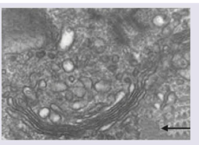
In this electron micrograph, identify the structure marked with arrow.

Which of the following proteins are not seen in the region marked in the image?

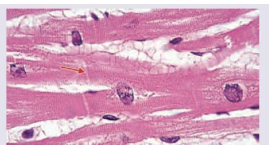
Which is correct about structures marked as "X" found in smooth muscle?

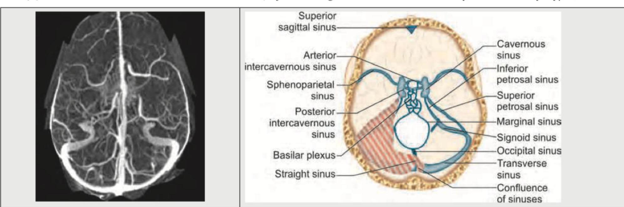
Identify the anatomical structure shown in the image.

Explanation: **G-banding (Giemsa banding)** is the gold standard and most frequently used technique for routine clinical cytogenetic analysis [1]. In this method, chromosomes are first treated with **Trypsin** (to partially digest proteins) and then stained with **Giemsa stain**. This produces a characteristic pattern of alternating light and dark bands: * **Dark bands (G-positive):** Represent AT-rich, gene-poor, heterochromatic regions that replicate late. * **Light bands (G-negative):** Represent GC-rich, gene-dense, euchromatic regions that replicate early. The stability and high resolution of these bands allow for the identification of specific chromosomes and the detection of structural abnormalities like deletions or translocations. **Analysis of Incorrect Options:** * **B. C-banding (Constitutive heterochromatin):** Specifically stains the centromeres and regions containing repetitive DNA (like 1q, 9q, 16q). It is not used for general screening. * **C. R-banding (Reverse banding):** Produces a pattern opposite to G-banding (dark bands are GC-rich). It is primarily used to study the distal ends (telomeres) of chromosomes which may be faint on G-banding. * **D. Q-banding (Quinacrine):** The first banding method developed; it uses fluorescent microscopy. It is less common today because the fluorescence fades quickly (photobleaching). **High-Yield Clinical Pearls for NEET-PG:** * **Karyotyping** is typically performed on cells arrested in **Metaphase** (using Colchicine) because chromosomes are most condensed [1]. * **Most common sample** for postnatal karyotyping: **Peripheral blood T-lymphocytes** (stimulated by Phytohemagglutinin). * **Resolution:** Standard G-banding identifies ~400–550 bands per haploid set; High-resolution banding (prophase/prometaphase) can identify up to 850+ bands [1].
Explanation: The Distal Convoluted Tubule (DCT) is a critical segment of the nephron located within the renal cortex. It begins at the macula densa (at the end of the thick ascending limb of Henle) and terminates by emptying into the collecting duct [2]. **Why 5 mm is correct:** In standard human anatomy, the DCT is significantly shorter and less convoluted than the Proximal Convoluted Tubule (PCT). While the PCT measures approximately 14–15 mm in length [1], the **DCT measures approximately 4.5 to 5 mm**. This shorter length and fewer microvilli are key histological features that distinguish it from the PCT under a microscope [1]. **Analysis of Incorrect Options:** * **A. 2 mm:** This is too short for the DCT; however, it is closer to the length of the *connecting tubule* that joins the DCT to the collecting duct. * **C. 8 mm & D. 12 mm:** These values are too high for the DCT. A length of 12–15 mm is characteristic of the **Proximal Convoluted Tubule (PCT)**, which is the longest and most bulky part of the nephron to facilitate bulk reabsorption [1]. **High-Yield Facts for NEET-PG:** * **Histology:** The DCT is lined by **simple cuboidal epithelium**. Unlike the PCT, it **lacks a prominent brush border** (fewer microvilli), making the lumen appear clearer and wider in cross-section [1]. * **Macula Densa:** The initial part of the DCT contains specialized cells called the macula densa, which act as chemoreceptors for sodium chloride and form part of the **Juxtaglomerular Apparatus (JGA)** [3]. * **Hormonal Action:** The late DCT is the primary site for facultative water reabsorption (via ADH) and sodium reabsorption/potassium secretion (via Aldosterone) [2]. * **Mnemonic:** "P" is for Proximal and **P**rolonged (15mm); "D" is for Distal and **D**iminished (5mm).
Explanation: **Explanation:** The correct answer is **D (It is a strict anaerobe)** because *Brucella* species are actually **strict aerobes**. They require oxygen for growth and do not ferment carbohydrates in conventional media. Some species, notably *Brucella abortus*, are capnophilic, requiring 5–10% $CO_2$ for initial isolation. **Analysis of Options:** * **Option A:** *Brucella* causes **Brucellosis**, clinically known as **Undulant fever** (due to the characteristic rising and falling temperature pattern), **Malta fever**, or **Mediterranean fever**, reflecting its geographical prevalence and historical discovery. * **Option B:** Morphologically, *Brucella* are small, non-motile, non-sporing, **Gram-negative coccobacilli**. They often appear as "fine grains of sand" under the microscope. * **Option C:** *Brucella* is highly susceptible to heat. **Pasteurization** of milk is the most effective public health measure to eliminate the transmission of bovine brucellosis to humans. **High-Yield Clinical Pearls for NEET-PG:** * **Zoonosis:** It is primarily a zoonotic disease transmitted via unpasteurized dairy or direct contact with infected animal tissues (placenta/secretions). * **Intracellular Pathogen:** It is a **facultative intracellular** organism, surviving within macrophages, which leads to chronicity and granuloma formation. * **Diagnosis:** The **Standard Agglutination Test (SAT)** is commonly used; a titer of 1:160 or more is significant. * **Culture:** **Castaneda’s medium** (biphasic medium) is the traditional method for blood culture to reduce the risk of laboratory-acquired infection. * **Treatment:** The WHO recommends **Doxycycline + Rifampicin** for 6 weeks.
Explanation: **Explanation:** **Hypersensitivity Vasculitis** (also known as Leukocytoclastic Vasculitis) is a type of small-vessel vasculitis typically mediated by **Type III hypersensitivity** (immune-complex deposition). 1. **Why Post-capillary Venules are Correct:** The **post-capillary venule** is the primary site of involvement because it is the segment of the microvasculature where blood flow is slowest and the endothelium is most responsive to inflammatory mediators. Immune complexes (antigen-antibody) circulate and deposit in these vessel walls, triggering the complement cascade. This leads to the recruitment of neutrophils, which release lysosomal enzymes, causing "leukocytoclasis" (nuclear debris/dust) and fibrinoid necrosis. 2. **Why Other Options are Incorrect:** * **Arterioles & Capillaries:** While small-vessel vasculitis can occasionally involve these, the classic histological hallmark and the predominant site of clinical manifestation (like palpable purpura) is the post-capillary venule. * **Medium-sized Arteries:** These are involved in conditions like **Polyarteritis Nodosa (PAN)** or **Kawasaki disease**. Hypersensitivity vasculitis, by definition, is a small-vessel disease and does not affect muscular arteries. **NEET-PG High-Yield Pearls:** * **Clinical Presentation:** The most common clinical sign is **palpable purpura**, usually found on dependent areas like the lower legs. * **Histopathology:** Look for **"nuclear dust"** (leukocytoclasis) and **fibrinoid necrosis** of the vessel wall. * **Triggers:** Often induced by drugs (penicillin, sulfa drugs), infections, or systemic autoimmune diseases. * **Classification:** It is categorized under **Small Vessel Vasculitis** (along with GPA, EGPA, and MPA), but unlike the ANCA-associated vasculitides, it is primarily immune-complex mediated.
Explanation: ***Apical*** - Tight junctions, also known as **zonula occludens**, are located at the **apical region** of polarized epithelial cells [1]. - They form the most **apical component** of the junctional complex, positioned at the apical-most part of the lateral cell membrane, just below the free apical surface [1]. - They are crucial for forming a **permeability barrier** that controls paracellular transport and maintains cell polarity by separating apical from basolateral membrane domains [2]. *Incorrect Apicolateral* - While tight junctions are technically at the interface between apical and lateral domains, "apicolateral" is **not standard anatomical terminology** used in medical textbooks. - The standard anatomical description places tight junctions at the **apical region** of epithelial cells. *Incorrect Basolateral* - The **basolateral domain** encompasses the lateral cell membrane (where adhesion junctions like desmosomes and communication junctions like gap junctions are located) and the basal membrane. - Tight junctions are positioned **above** these other junctional complexes, at the apical-most position [1]. *Incorrect Basal* - The basal surface rests on the **basement membrane**. - The characteristic junction here is the **hemidesmosome**, which anchors the cell to the underlying extracellular matrix, not to seal adjacent cells [1].
Explanation: ***X=H zone, Y=A band*** - **X** points to the central region of the A band, visible only in a relaxed sarcomere, which is called the **H zone**, containing only thick myosin filaments. - **Y** encompasses the entire length of the thick filaments, including the regions where they overlap with thin filaments, defining the **A band**. *X=A band, Y=H band* - This is incorrect because X specifically indicates the central, lighter region within the A band, which is the H zone. - Y points to the entire segment occupied by the thick filaments, which is the A band. *X=Z line, Y=M line* - The **Z line** marks the boundaries of a sarcomere, anchoring the thin (actin) filaments, and is not indicated by X. - The **M line** is the central line within the H zone that anchors the thick (myosin) filaments, and is not indicated by Y. *X=M line, Y=Z line* - As explained, X indicates the **H zone**, which is a broader region than the M line. - Y indicates the **A band**, and not the Z line; the Z line is located at the ends of the sarcomere.
Explanation: ***Smooth endoplasmic reticulum*** - The image shows a network of **tubular structures** that are interconnected and lack ribosomes, which is characteristic of the smooth endoplasmic reticulum. - The smooth endoplasmic reticulum is involved in various metabolic processes, including **lipid synthesis**, detoxification, and calcium storage. *Trans Golgi network* - The trans Golgi network consists of **flattened sacs (cisternae)** and is the exit face of the Golgi apparatus, where proteins are sorted and packaged into vesicles. - It would appear as a series of stacked, flattened membranes, often associated with budding vesicles, which is not what the arrow indicates. *Cis Golgi network* - The cis Golgi network is the entry face of the Golgi apparatus, receiving proteins from the ER. It also consists of **flattened cisternae**. - It is located closest to the endoplasmic reticulum and looks like flattened sacs rather than a tubular network. *Medial Golgi network* - The medial Golgi network comprises the cisternae located between the cis and trans faces, involved in further processing and modification of proteins. - Like the cis and trans networks, it is composed of **flattened, stacked cisternae**, distinct from the tubular appearance of the structure pointed to by the arrow.
Explanation: ***Zona occludens*** - The image shows **cardiac muscle** tissue, and the arrow points to an **intercalated disc**. - Intercalated discs are primarily composed of **fascia adherens**, **maculae adherentes (desmosomes)**, and **gap junctions (connexons)**, but not tight junctions (zona occludens). *Macula adherens* - **Maculae adherentes**, also known as **desmosomes**, are abundant in intercalated discs. - They provide **strong adhesion** between cardiac muscle cells and are crucial for resisting mechanical stress. *Fascia adherens* - **Fascia adherens** are the most extensive type of junction in the transverse portion of the intercalated disc. - They anchor the **actin filaments** of the terminal sarcomeres to the plasma membrane. *Connexions* - **Connexons** are the structural proteins that form **gap junctions**. - Gap junctions in intercalated discs allow for the rapid **passage of ions** and small molecules, facilitating electrical coupling and coordinated contraction.
Explanation: ***Dense bodies*** - The structures marked "X" are **dense bodies**, which are analogous to **Z-discs** in skeletal muscle, serving as attachment points for **actin filaments**. - They are crucial for transmitting the contractile force generated by the **actin-myosin bundles** to the cell membrane, leading to the characteristic corkscrew-like contraction of smooth muscle cells. *A band* - The **A band** is a region found in **striated muscle** (skeletal and cardiac muscle) where thick and thin filaments overlap, corresponding to the length of the myosin filaments. - Smooth muscle lacks the organized sarcomeric structure, and thus, **A bands** are not present. *Calmodulin* - **Calmodulin** is a calcium-binding protein that plays a key role in smooth muscle contraction by activating **myosin light chain kinase (MLCK)**. - However, it is a soluble protein involved in signaling, not a structural component like the dense bodies shown in the image. *H band* - The **H band** is located within the **A band** of a **sarcomere** in striated muscle, representing the central region where only thick (myosin) filaments are present. - Similar to the **A band**, the **H band** is a feature of striated muscle and is not found in the unstriated smooth muscle cells.
Explanation: ***Transverse sinus*** - The image shows a venogram with the highlighted structure corresponding to the **transverse sinus**, a large dural venous sinus located in the posterior cranial fossa - The transverse sinus extends **laterally from the confluence of sinuses** along the attached margin of the tentorium cerebelli - It curves anteroinferiorly to become the **sigmoid sinus** at the posterolateral corner of the petrous temporal bone - Drains blood from the superior sagittal sinus and straight sinus via the confluence of sinuses *Incorrect: Superior sagittal sinus* - The superior sagittal sinus runs in the **midline along the superior border** of the falx cerebri - On venogram, it appears as a **midline vertical structure**, not the lateral structure shown - Drains into the confluence of sinuses but has a different anatomical course *Incorrect: Sigmoid sinus* - The sigmoid sinus is the **continuation of the transverse sinus** - It follows an **S-shaped course** descending to the jugular foramen - Located more **anteroinferiorly** than the structure shown in the image *Incorrect: Straight sinus* - The straight sinus runs **posteriorly in the midline** along the junction of falx cerebri and tentorium cerebelli - Drains into the confluence of sinuses but is **not laterally positioned** like the structure shown - On venogram, it appears more posterior and vertical compared to the transverse sinus
Cellular Ultrastructure
Practice Questions
Microscopic Anatomy of Epithelial Tissues
Practice Questions
Microscopic Anatomy of Connective Tissues
Practice Questions
Microscopic Anatomy of Muscle Tissues
Practice Questions
Microscopic Anatomy of Nervous Tissues
Practice Questions
Microscopic Anatomy of Blood and Immune System
Practice Questions
Microscopic Anatomy of Endocrine Glands
Practice Questions
Microscopic Anatomy of Digestive System
Practice Questions
Microscopic Anatomy of Respiratory System
Practice Questions
Microscopic Anatomy of Urinary System
Practice Questions
Microscopic Anatomy of Reproductive System
Practice Questions
Techniques in Microscopic Anatomy
Practice Questions
Get full access to all questions, explanations, and performance tracking.
Start For Free