
Microscopic Anatomy of Muscle Tissues — MCQs

All of the following are true about skeletal muscle except?
What is the anatomical arrangement of fibers in the masseter muscle that contributes to its strength?
Arrange the following parts of sarcomere from periphery to center. 1. Z line 2. M line 3. A band 4. H zone
A 4-year-old boy is brought to the physician by his parents due to frequent falls, inability to jump, and easy fatigue. Physical examination reveals weakness in the pelvic and shoulder girdles, as well as enlargement of the child's calf muscles. The serum level of creatine kinase is elevated. A biopsy of calf muscle reveals marked variation in size and shape of muscle fibers, with foci of muscle fiber necrosis, myophagocytosis, regenerating fibers, and fibrosis. Molecular diagnostic assays performed on muscle biopsy from the patient would show alterations in the length of the primary transcript for which muscle-associated protein?
Single-unit smooth muscles are seen in which of the following?
Which of the following are supportive proteins?
What is a key difference between smooth muscle and skeletal muscle physiology?
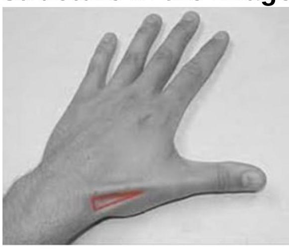
The image shows a highlighted region on the dorsal aspect of the hand (anatomical snuffbox). Which of the following anatomical structures form the boundaries or floor of this region?
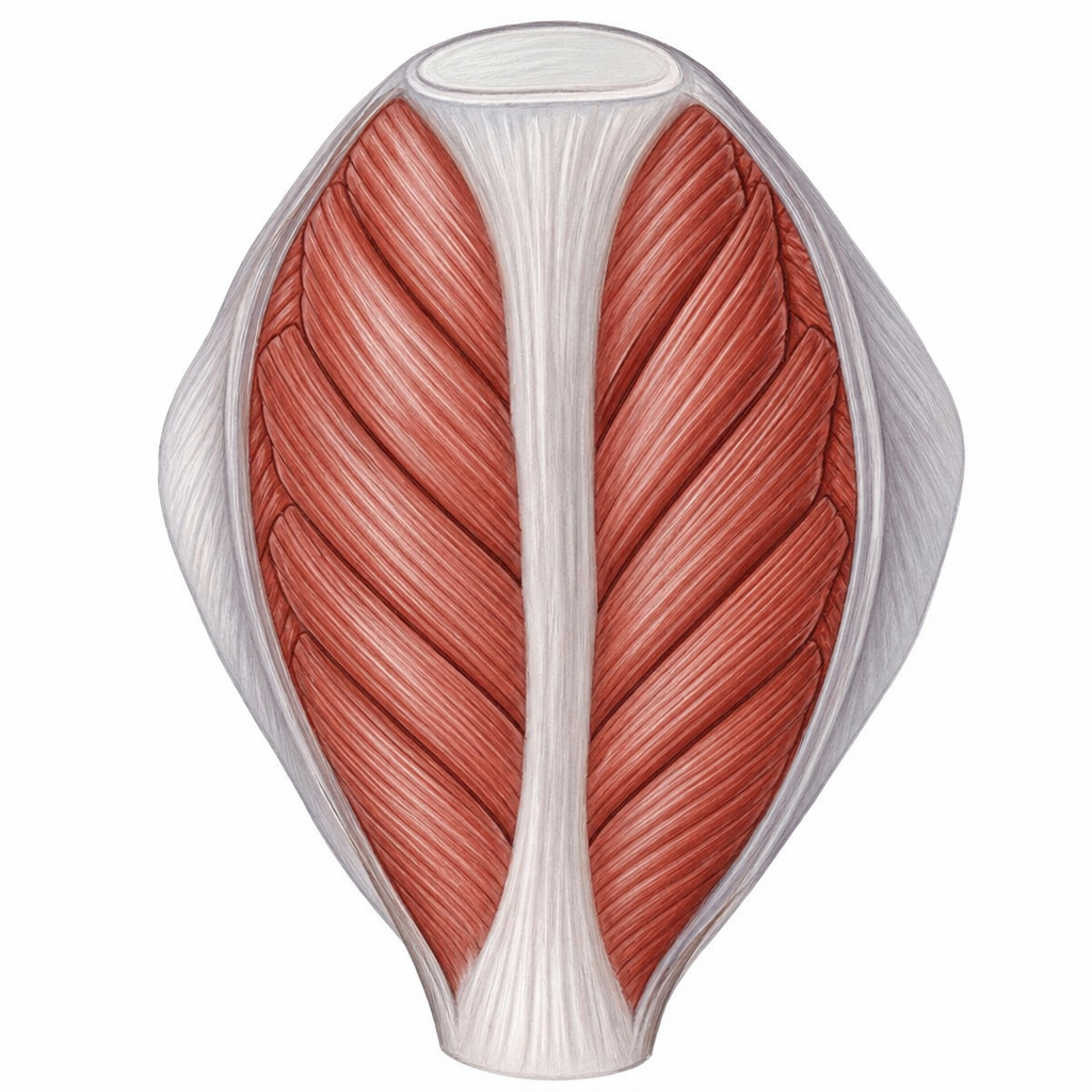
Identify the type of muscle shown in the image.
The lateral boundary of the cubital fossa is formed by
Want unlimited practice?
Get full access to all questions, explanations, and performance tracking.
Scan to download app