Froment's sign (book test) is used to assess the function of adductor pollicis. Which of the following nerves supplies this muscle?
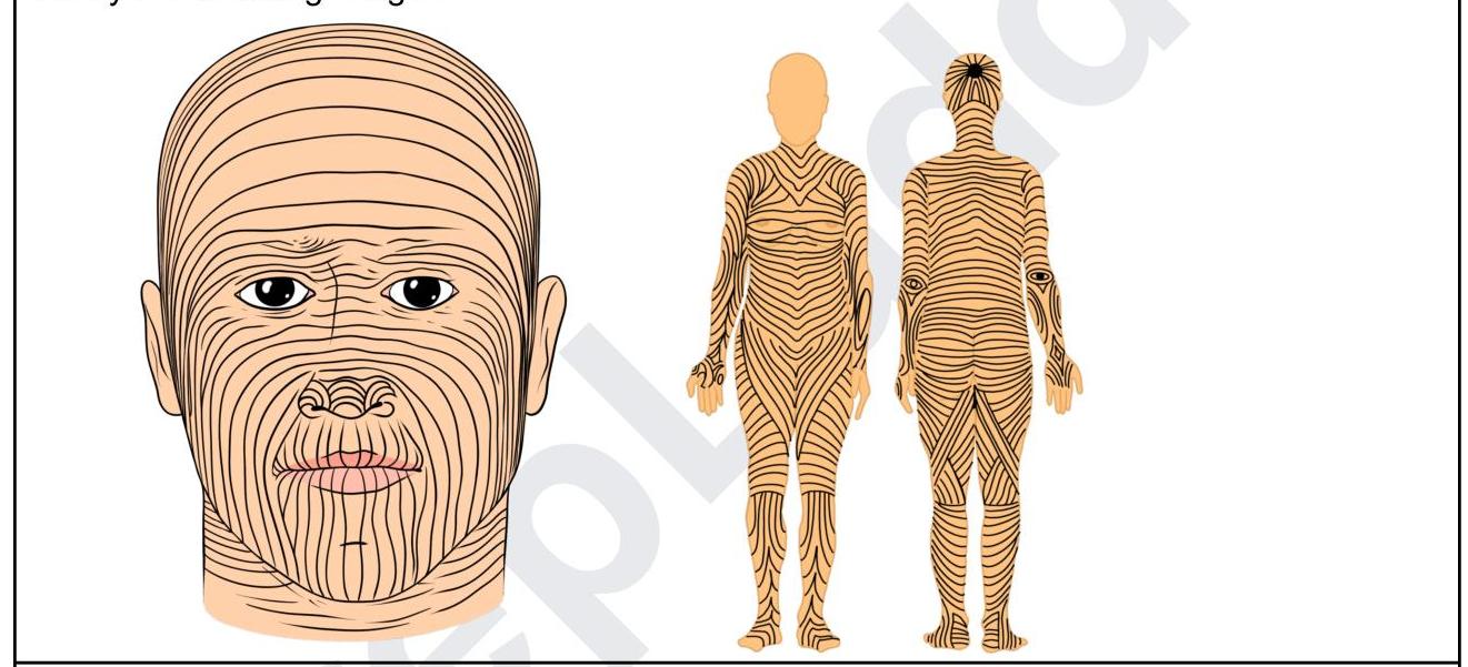
Identify the lines shown in the following image:

A patient after a road traffic accident presents to the emergency room with difficulty in swallowing and slurred speech. Investigations reveal fractures in the occipitotemporal region. Which of the following areas should be tested in order to find the nerve which is involved?
A patient who has taken the first COVID vaccine comes for the second dose. An astute nurse noticed that the shoulder was flabby, flat, and was asymmetrical. There was an associated loss of contour of the shoulder joint. Injury to which of the structures might have resulted and was avoidable?
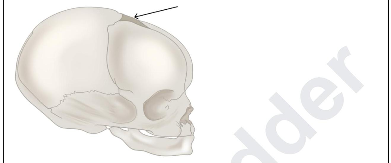
A 6-month-old boy was brought to the casualty with seizures. The pediatrician tries to do CSF sampling. What are the structures punctured by the pediatrician while piercing through the marked structure?

During a thyroidectomy, a surgeon must carefully identify and preserve the parathyroid glands. These glands are most commonly located posterior to which part of the thyroid gland?
An MRI of a patient with low back pain reveals compression of the L5 nerve root. Which of the following muscles would most likely show weakness during physical examination?
A 45-year-old woman undergoes a modified radical mastectomy for breast cancer. Following the procedure, she experiences numbness in the medial aspect of her upper arm. Which of the following nerves was most likely injured during the surgery?
In which location is a transplanted kidney typically placed?
Winging of scapula is due to paralysis of
Explanation: The adductor pollicis muscle is primarily innervated by the deep branch of the ulnar nerve [1]. This muscle is crucial for thumb adduction, and its function is assessed by the Froment's sign (often referred to as the book test) to detect ulnar nerve palsy. The radial nerve primarily innervates the muscles of the posterior compartment of the arm and forearm, responsible for wrist and finger extension [1]. It does not supply any intrinsic muscles of the hand, including the adductor pollicis [1]. The posterior interosseous nerve is a terminal branch of the deep radial nerve, supplying most of the extensors in the forearm [1]. It does not innervate any of the intrinsic hand muscles. The median nerve innervates most of the flexors of the forearm and the thenar muscles of the hand (abductor pollicis brevis, opponens pollicis, and superficial head of flexor pollicis brevis) [1]. It does not supply the adductor pollicis muscle; median nerve palsy would affect thumb opposition and abduction, not adduction [1].
Explanation: ***Langer's lines*** - The image displays lines that represent the **natural orientation of collagen fibers** within the human skin, which are known as Langer's lines (also called cleavage lines). - Making surgical incisions **parallel to these lines** can result in better wound healing and less scarring. - Named after **Karl Langer**, an Austrian anatomist who described these lines in 1861. *Hinderer's lines* - While **Hinderer** described relaxed skin tension lines (RSTLs) used in plastic surgery, these are **different from Langer's lines**. - The image shows Langer's lines specifically, which are based on **collagen fiber orientation**, not relaxed skin tension. *Dermatomes* - **Dermatomes** are areas of skin mainly supplied by a single **spinal nerve root**. - They represent **neurologic segments** and do not correspond to the collagen fiber orientation shown in the image. *Blaschko's lines* - **Blaschko's lines** are invisible lines of skin cell migration that become visible in certain **genetic or acquired dermatological conditions**. - They represent a **mosaic pattern** due to different cell populations and are distinctly different from the structural collagen lines shown.
Explanation: ***Posterior one-third of tongue*** - This symptom complex of **dysphagia** (difficulty swallowing) and **dysarthria** (slurred speech) after trauma to the occipitotemporal region is highly suggestive of damage to **Cranial Nerves IX (Glossopharyngeal)** and **X (Vagus)**. - The **Glossopharyngeal nerve (CN IX)** supplies general and special sensation (taste) to the **posterior one-third of the tongue** [1]. *Anterior two-thirds of tongue* - The **facial nerve (CN VII)** is responsible for taste sensation from the **anterior two-thirds of the tongue** [1]. - General sensation from the anterior two-thirds of the tongue is supplied by the **trigeminal nerve (CN V)** via the lingual nerve. *Hard palate* - Sensation to the **hard palate** is primarily supplied by branches of the **trigeminal nerve (CN V)**, specifically the greater palatine and nasopalatine nerves. - Damage to these nerves would primarily affect sensation in the palate, not cause dysphagia and dysarthria. *Soft palate* - The **vagus nerve (CN X)** is responsible for motor innervation to most muscles of the **soft palate**, allowing for its elevation during swallowing and speech. - While soft palate dysfunction can contribute to dysphagia and dysarthria, directly testing sensation here would be less specific than testing the posterior tongue for Glossopharyngeal involvement.
Explanation: ***Deltoid muscle*** - The **deltoid muscle** is the principal muscle that gives the shoulder its rounded contour. Damage to or atrophy of the deltoid can lead to a **flat, flabby, and asymmetrical shoulder**. - Improper vaccine administration, such as injecting too high or too deep, can directly injure the deltoid muscle, leading to inflammation (**SIRVA - Shoulder Injury Related to Vaccine Administration**) or even deltoid atrophy, which would cause the observed lack of contour. *Supraspinatus muscle* - The **supraspinatus** is primarily involved in the initial **abduction** of the arm and stabilization of the shoulder joint, but it does not significantly contribute to the visible external contour of the shoulder. - Injury to the supraspinatus mainly causes **pain and weakness** with abduction, rather than a visible change in shoulder shape. *Infraspinatus muscle* - The **infraspinatus** is a rotator cuff muscle primarily responsible for **external rotation** of the arm. - Injury to this muscle would cause weakness in external rotation and potentially posterior shoulder pain, but not the noticeable loss of shoulder contour described. *Teres minor muscle* - The **teres minor** is also a rotator cuff muscle assisting in **external rotation** and stabilization of the humeral head. - Similar to the infraspinatus, its injury would impair external rotation and cause posterior shoulder pain, but it doesn't define the overall shape of the shoulder.
Explanation: ***Scalp, dura, arachnoid*** - The image shows a needle piercing through the **anterior fontanelle**, which allows direct access to the intracranial space. - When accessing the **subarachnoid space** via the fontanelle, the needle would penetrate the overlying **scalp**, then the outer and inner layers of the **dura mater**, and finally the **arachnoid mater** before reaching the cerebrospinal fluid. *Scalp, epicranium, endocranium and dura* - The terms **epicranium** and **endocranium** refer to layers associated with the bone itself, which is largely absent as a solid structure at the fontanelle in an infant. - CSF sampling through the fontanelle bypasses the need to penetrate mature bone layers like epicranium and endocranium. *Scalp synchondral membrane, dura, arachnoid* - A **synchondral membrane** is found between bones that are joined by cartilage, such as at the base of the skull, not typically within the fontanelle itself. - The primary layers to be penetrated at the fontanelle are the soft tissues of the scalp and the meningeal layers. *Pericranium, dura, arachnoid* - The **pericranium** is the dense connective tissue layer that covers the outer surface of the bones of the skull. - While present, it's considered part of the overall scalp layers and is not a separate primary penetration layer in the same context as the meninges for CSF sampling.
Explanation: Detailed anatomical knowledge is crucial during thyroidectomy to ensure preservation of vital structures [1]. ***Inferior poles*** - The **inferior parathyroid glands** (parathyroid IV) are most commonly located posterior to the **inferior poles** of the thyroid gland [1]. - While they are more variable in position than superior glands and can descend into the thymus or mediastinum, the **most common location** is still posterior to the inferior poles [1]. - During thyroidectomy, these glands are frequently encountered in the inferior pole region and must be carefully preserved [1]. *Superior poles* - The **superior parathyroid glands** (parathyroid III) are typically found at the **middle-to-upper third** of the thyroid, near the cricothyroid junction, rather than directly at the superior poles. - While their position is more constant than inferior glands, they are not specifically located at the superior poles themselves. *Pyramidal lobe* - The **pyramidal lobe** is an embryological remnant extending superiorly from the thyroid isthmus. - It is not associated with parathyroid gland location, as parathyroids are distinct endocrine structures located on the posterior thyroid surface. *Middle third* - The **superior parathyroid glands** are often found near the middle third of the thyroid posteriorly. - However, when considering all four parathyroid glands (both superior and inferior pairs), the **inferior glands** at the inferior poles represent the most common overall location pattern.
Explanation: ***Tibialis anterior*** - The **L5 nerve root** primarily innervates muscles responsible for **dorsiflexion** of the foot, with the **tibialis anterior** being the primary dorsiflexor. - Weakness of the tibialis anterior would manifest as difficulty lifting the front of the foot, potentially leading to a **foot drop** gait. *Tibialis posterior* - The **tibialis posterior** is primarily innervated by the **tibial nerve** (S1-S2) and is responsible for **plantarflexion** and **inversion** of the foot. - Weakness in this muscle would not be the most likely presentation of L5 nerve root compression. *Gastrocnemius* - The **gastrocnemius** muscle is primarily innervated by the **tibial nerve** (S1-S2) and is a powerful **plantarflexor** of the foot. - Weakness in this muscle would indicate an S1 or S2 nerve root issue, not typically L5. *Quadriceps femoris* - The **quadriceps femoris** is innervated by the **femoral nerve**, predominantly originating from the **L2, L3, and L4 nerve roots**. - Weakness would manifest as difficulty extending the knee, which is not characteristic of L5 compression.
Explanation: ***Intercostobrachial nerve*** - The **intercostobrachial nerve** provides sensory innervation to the **medial aspect of the upper arm** and is vulnerable to injury during **axillary dissection** in a modified radical mastectomy [1]. - Injury typically results in **numbness** or **paresthesia** in this specific dermatomal distribution [1]. *Musculocutaneous nerve* - The **musculocutaneous nerve** innervates the muscles of the **anterior compartment of the arm** (e.g., biceps brachii) and provides sensation to the **lateral forearm**. - Damage would primarily affect **forearm sensation** and arm flexion, not medial upper arm sensation. *Thoracodorsal nerve* - The **thoracodorsal nerve** innervates the **latissimus dorsi muscle**, a large muscle of the back and shoulder [1]. - Injury would lead to **weakness in adduction, extension, and internal rotation** of the arm, with no sensory deficit in the upper arm [1]. *Long thoracic nerve* - The **long thoracic nerve** innervates the **serratus anterior muscle**, which stabilizes the scapula and allows for arm abduction above 90 degrees. - Injury results in **"winged scapula,"** making it difficult to raise the arm overhead, without sensory loss in the upper arm.
Explanation: ***Iliac fossa*** - The **iliac fossa** is the standard site for kidney transplantation due to its accessibility and the proximity of suitable blood vessels (iliac artery and vein) for anastomosis. [1] - Placing the kidney here allows for easier access for potential biopsies and monitoring. *Retroperitoneal space* - The patient's native kidneys are located in the **retroperitoneal space**, but a transplanted kidney is typically placed in a more accessible anterior location. - Transplanting a kidney into the retroperitoneal space would be a more complex and invasive procedure due to the depth and surrounding structures. *Hypogastric region* - While the iliac fossa is part of the broader hypogastric region, the term **hypogastric region** is less specific for the precise anatomical location of kidney transplantation. - The iliac fossa specifically refers to the concave surface of the ilium, which provides a suitable bed for the donated kidney. *Kidney fossa* - **Kidney fossa** is not a formally recognized anatomical term for a specific site of kidney transplantation. - The term "fossa" describes a depression or hollow, but in the context of transplantation, the iliac fossa is the correct anatomical landmark used.
Explanation: ***Serratus anterior*** - The **serratus anterior muscle** is responsible for **protraction and rotation of the scapula**, holding it close to the thoracic wall. - Paralysis of this muscle, often due to injury to the **long thoracic nerve**, causes the **medial border of the scapula** to protrude posteriorly, a condition known as **medial scapular winging**. - This is the **classic and most common cause** of scapular winging. *Rhomboid major* - The rhomboid major muscle primarily performs **retraction and downward rotation of the scapula**. - Paralysis of this muscle would lead to the scapula being displaced laterally and superiorly, not winging. *Trapezius* - The trapezius muscle has multiple actions, including **elevating, depressing, retracting, and rotating the scapula**. - Paralysis of the trapezius (e.g., due to **accessory nerve damage**) can cause **lateral scapular winging** where the inferior angle protrudes, along with shoulder drooping and difficulty shrugging. - However, **serratus anterior paralysis** is the classic answer for scapular winging in exam contexts. *Levator scapulae* - The levator scapulae muscle is primarily involved in **elevating and downwardly rotating the scapula**. - Dysfunction of this muscle would impair shoulder elevation but would not be the direct cause of scapular winging.
Anatomical Basis of Common Clinical Conditions
Practice Questions
Surgical Anatomy
Practice Questions
Anatomical Basis of Trauma
Practice Questions
Anatomical Aspects of Infections
Practice Questions
Anatomical Considerations in Regional Anesthesia
Practice Questions
Anatomical Basis of Physical Examination
Practice Questions
Clinical Correlations in Neuroanatomy
Practice Questions
Anatomical Approaches in Minimally Invasive Procedures
Practice Questions
Imaging Correlations in Clinical Anatomy
Practice Questions
Anatomical Variations of Clinical Importance
Practice Questions
Get full access to all questions, explanations, and performance tracking.
Start For Free