Name the test being performed.

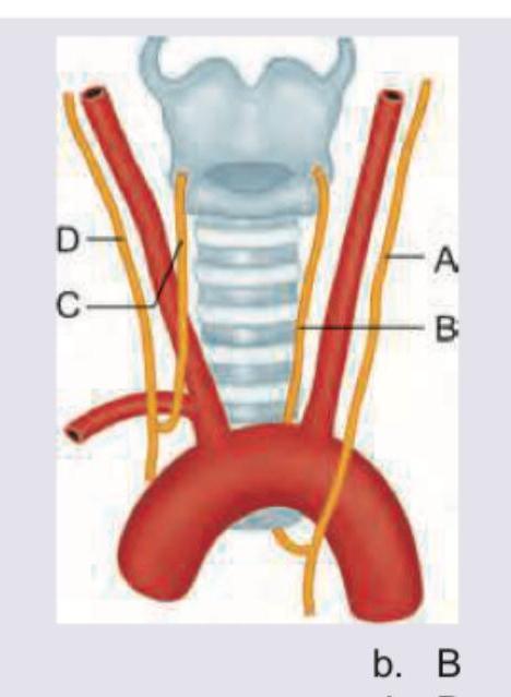
A 15-year-old child with rheumatic heart disease is having hoarseness of voice. Mark the nerve involved in the diagram shown below:

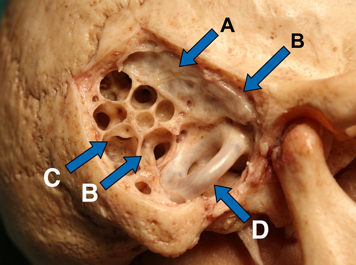
An intra-operative photograph of cortical mastoidectomy is shown. Identify the lateral semi-circular canal. (AIIMS Nov 2017)

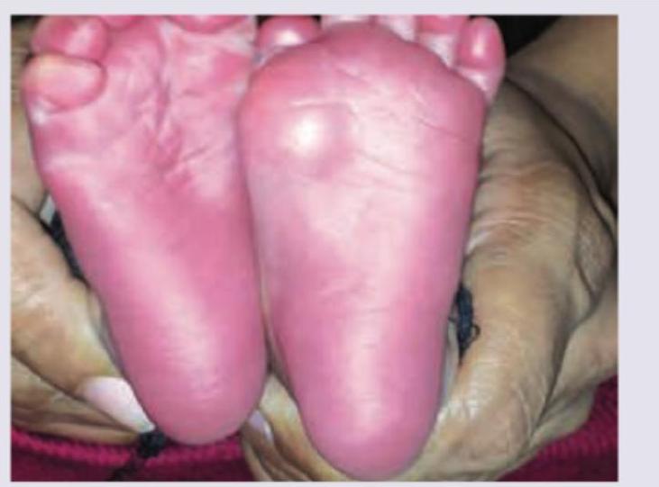
All are seen in this girl child except:

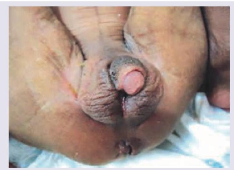
What investigations and management are indicated in the condition shown in the image?

A child is brought to your OPD with rashes as shown below along with fever. Diagnosis is:

What is the location of Erb's point during auscultation?
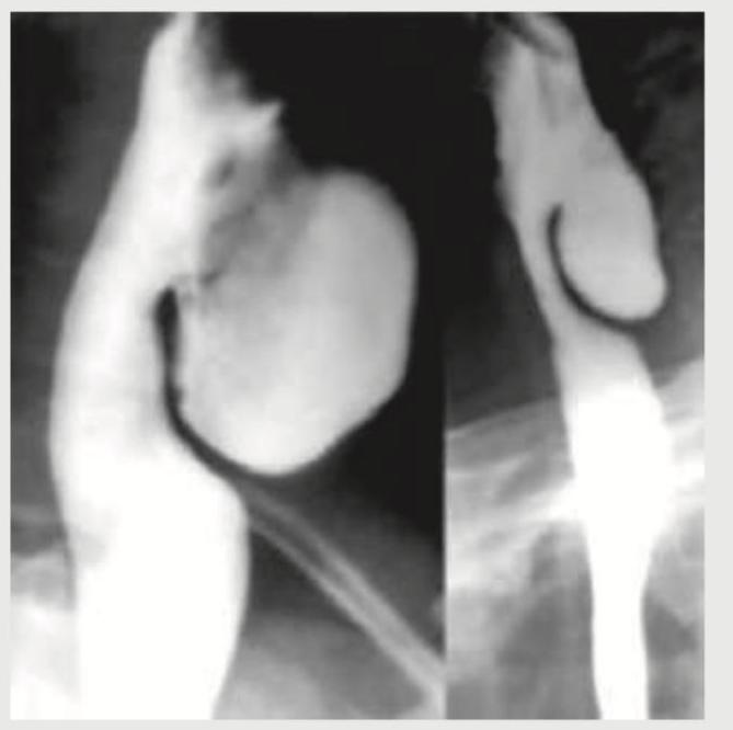
A 58-year-old male patient presents with halitosis, mild dysphagia and regurgitation of previous day food. Barium study is performed. Where is the location of this presentation?

Which of the statements regarding Calot's triangle are correct? 1. Common hepatic duct forms the medial boundary of the Calot's triangle 2. Inferior surface of the right lobe of the liver forms the superior boundary of Calot's triangle 3. Right hepatic artery is usually found as a content of the Calot's triangle 4. Cystic duct and medial border of gall bladder forms the lateral border of Calot's triangle Select the correct answer using the code given below:
Ptosis in Horner syndrome is due to paralysis of which muscle?
Explanation: ***Kirby-Bauer test*** - The image displays a **Kirby-Bauer disk diffusion test**, evident by the **antibiotic-impregnated paper disks** placed on an agar plate inoculated with bacteria. - The clear areas around the disks are **zones of inhibition**, indicating the antimicrobial activity of the respective antibiotics against the test organism. *E-test* - An E-test involves a **plastic strip containing a gradient of antibiotic concentration**. - It provides a **minimum inhibitory concentration (MIC)** value, which is not depicted in this image. *CLO test* - The CLO (Campylobacter-like organism) test is a rapid diagnostic test for the presence of **Helicobacter pylori** in gastric biopsy samples. - It detects **urease activity**, leading to a color change, which is unrelated to the image shown. *Levaditi test* - The Levaditi test is a historical **silver-staining method** used to visualize **spirochetes**, particularly *Treponema pallidum*, in tissue samples. - It is a histological staining technique, not an antimicrobial sensitivity test on an agar plate.
Explanation: ***Correct Option B*** - The image shows the **left recurrent laryngeal nerve** (indicated by label B) looping around the **aortic arch** and ascending towards the larynx. - In rheumatic heart disease, severe **mitral stenosis** can lead to enlargement of the **left atrium**, compressing the left recurrent laryngeal nerve against the aortic arch, resulting in **hoarseness of voice (Ortner's syndrome)**. *Incorrect Option A* - This structure (A) represents the **right recurrent laryngeal nerve**, which typically loops around the **right subclavian artery** and is not usually implicated in Ortner's syndrome due to left atrial enlargement. - While damage to this nerve can also cause hoarsiness, it would not be related to the pathophysiology of cardiac enlargement in rheumatic heart disease. *Incorrect Option C* - This structure (C) represents the **vagus nerve** (cranial nerve X) in the neck, from which the recurrent laryngeal nerves branch. - Direct compression of the vagus nerve in this location is less common as a cause of isolated hoarseness related to cardiac pathology compared to the recurrent laryngeal nerve. *Incorrect Option D* - This structure (D) represents a major blood vessel in the neck, likely the **left common carotid artery** or **left internal jugular vein**, both of which are not directly involved in phonation or compressed by atrial enlargement in a way that causes hoarseness. - These vessels are primarily involved in blood supply to and drainage from the head and neck.
Explanation: ***Correct Option D*** - The diagram shows a simplified view of the temporal bone anatomy post-mastoidectomy, and 'D' clearly points to the position of the **lateral semicircular canal**. - During cortical mastoidectomy, the **lateral semicircular canal** is typically identified in the superior and posterior wall of the mastoid cavity, just above the aditus and facial nerve. - This is a critical landmark during mastoid surgery as injury to it can cause vertigo and hearing loss. *Incorrect Options A, B, and C* - These labels point to other anatomical structures visible in the mastoid cavity during cortical mastoidectomy, such as the tegmen tympani, facial nerve, sigmoid sinus, or other osseous landmarks. - Proper identification of the lateral semicircular canal is essential to avoid iatrogenic injury during surgery.
Explanation: The image shows a newborn with **ambiguous genitalia**, which is highly suggestive of **Congenital Adrenal Hyperplasia (CAH)** due to **21-hydroxylase deficiency** in a **genetic female (XX)** infant. The clitoris is enlarged to resemble a penis, and the labia are fused, appearing like a scrotum. In CAH, there's impaired cortisol synthesis, leading to increased ACTH and accumulation of androgen precursors. ***Girl children are more likely to die than boys*** - This statement is **FALSE** and is the correct answer to this "except" question. - In **genetic females (XX)** with CAH, the **ambiguous genitalia are recognized at birth**, leading to **earlier diagnosis and prompt management**. - In contrast, **genetic males (XY)** with CAH have **normal-appearing external genitalia** at birth, so their diagnosis is often **delayed until they present with a salt-wasting crisis** in the first few weeks of life. - Therefore, **boys are actually MORE likely to die than girls** in CAH, making the given statement incorrect. *Blood levels of 17-keto-steroids are markedly elevated* - In classic 21-hydroxylase deficiency, there is an impairment in the conversion of 17-hydroxyprogesterone to 11-deoxycortisol. - This leads to **accumulation of androgen precursors** such as DHEA and androstenedione. - **17-ketosteroids (metabolites of these androgens) are significantly elevated** in CAH, making this a true finding. *Hyponatremia and hyperkalemia* - In **salt-wasting CAH**, the deficiency of **aldosterone** leads to renal salt loss and impaired potassium excretion. - This results in **hyponatremia and hyperkalemia**, which are common and expected findings. - These electrolyte imbalances can be life-threatening and constitute an adrenal crisis. *Metabolic acidosis* - **Salt-wasting CAH** with severe dehydration and hypovolemic shock can lead to **metabolic acidosis** due to lactic acidosis from poor tissue perfusion. - **Hyperkalemia** itself can also contribute to metabolic acidosis. - While some texts mention metabolic alkalosis from volume contraction, in acute salt-wasting crisis, metabolic acidosis is commonly observed.
Explanation: ***Hematocrit and partial exchange transfusion with saline*** - The image shows a neonate with **plethora** (reddish-purple skin discoloration), characteristic of **polycythemia** (venous hematocrit >65%). - **Hematocrit** (venous sample) is the primary investigation to confirm polycythemia. - **Partial exchange transfusion with normal saline** is the **standard treatment** for symptomatic polycythemia, effectively reducing hematocrit from ~70% to ~55% while maintaining blood volume and improving blood flow. - The procedure reduces blood viscosity and prevents complications like hypoglycemia, seizures, renal vein thrombosis, and necrotizing enterocolitis. *Hematocrit and double volume exchange transfusion* - While **hematocrit** is the correct investigation, **double volume exchange transfusion** is NOT indicated for polycythemia. - Double volume exchange (replacing 2× blood volume) is reserved for **severe hemolytic disease with hyperbilirubinemia** (e.g., Rh/ABO incompatibility), not for polycythemia management. - Polycythemia requires only **partial exchange** to reduce hematocrit, not complete blood replacement. *Serum bilirubin and double volume exchange transfusion* - **Serum bilirubin** is not the primary investigation for polycythemia; the clinical picture suggests hyperviscosity, not jaundice. - This combination would be appropriate for severe **hemolytic jaundice**, not polycythemia. - Polycythemia is diagnosed by **elevated hematocrit**, not bilirubin levels. *Serum bilirubin and partial exchange transfusion with saline* - **Serum bilirubin** is not the initial investigation for polycythemia; **hematocrit** is required for diagnosis. - While partial exchange is the correct treatment approach, it must be guided by hematocrit values, not bilirubin levels.
Explanation: ***Meningococcemia*** - The image shows a **purpuric rash** with some confluent lesions, which are highly characteristic of **meningococcemia**, especially in a child with fever. - This rash is caused by **endotoxin-mediated vascular damage**, leading to petechiae and purpura that do not blanch under pressure. *Dengue haemorrhagic fever* - While dengue can cause petechiae and purpuric rashes, it typically presents with a more generalized rash, bleeding manifestations like **epistaxis** or **gum bleeding**, and often a history of mosquito exposure. - The rash in dengue is often described as an **"islands of white in a sea of red"** pattern, which is distinct from the more widespread, darker purpuric lesions seen in the image. *Leptospirosis* - Leptospirosis can manifest with a rash, but it is typically **maculopapular** or **erythematous**, sometimes appearing petechial. However, it rarely presents with the extensive, dark purpuric lesions shown, which are indicative of widespread microvascular damage. - Other classic features include **conjunctival suffusion**, myalgia, and severe headache, often following exposure to contaminated water or animal urine. *Scrub typhus* - Scrub typhus typically presents with a **maculopapular rash** that may become generalized, often sparing the face, palms, and soles. A characteristic **eschar** at the site of the chigger bite is a key diagnostic feature. - While petechiae can occur, the widespread, dark purpuric lesions seen in the image are not typical for scrub typhus.
Explanation: ***C=3rd intercostal space left parasternal line*** - **Erb's point** is a classic auscultation area located at the **third intercostal space** just to the left of the sternum. - While not corresponding to a specific single valve, it is where the sounds of **aortic** and **pulmonary regurgitation** can often be heard. *A=2nd intercostal space right parasternal line* - This location corresponds to the **aortic area**, primarily used for listening to the **aortic valve**. - It's optimal for detecting **aortic stenosis** and accentuation of the second heart sound (S2) due to aortic closure. *B=2nd intercostal space left parasternal line* - This is the **pulmonary area**, used for auscultating the **pulmonary valve**. - It is best for hearing murmurs related to **pulmonic stenosis** and **pulmonary hypertension**. *D=5th intercostal space left parasternal line* - This area is typically the **mitral area (apex)**, or occasionally just medial to it, for listening to the **mitral valve**. - It is optimal for assessing **mitral stenosis** and **regurgitation**, as well as the point of maximal impulse (PMI).
Explanation: ***Posterior segment of the esophagus along inferior pharyngeal constrictor muscle*** - The symptoms of **halitosis**, **dysphagia**, and **regurgitation of undigested food** are classic for a **Zenker's diverticulum**, which is a **pharyngoesophageal pouch**. - This diverticulum forms due to herniation of the pharyngeal mucosa through a weak point (Killian's triangle) in the **posterior pharyngeal wall**, specifically above the cricopharyngeus muscle (part of the **inferior pharyngeal constrictor**). *Anterior segment of the esophagus along superior pharyngeal constrictor muscle* - **Zenker's diverticulum** is located posteriorly, not anteriorly. - It arises in relation to the **inferior pharyngeal constrictor muscle**, not the superior pharyngeal constrictor. *Posterior segment of the esophagus along superior pharyngeal constrictor muscle* - While Zenker's diverticulum is **posterior**, it is associated with the **inferior pharyngeal constrictor muscle** (specifically the cricopharyngeus muscle in Killian's triangle), not the superior pharyngeal constrictor. - Herniation occurs above the **cricopharyngeus muscle**, which is the lowermost part of the inferior pharyngeal constrictor. *Anterior segment of the esophagus along inferior pharyngeal constrictor muscle* - **Zenker's diverticulum** is a **posterior** herniation, not an anterior one. - Although it involves the **inferior pharyngeal constrictor muscle**, its posterior location is a defining characteristic.
Explanation: ***1, 2 and 4*** - The **common hepatic duct** indeed forms the medial boundary, the **inferior surface of the right lobe of the liver** forms the superior boundary, and the **cystic duct along with the medial border of the gallbladder** forms the lateral boundary of Calot's triangle. - The **cystic artery** is typically a content of Calot's triangle [1], not the right hepatic artery. *1, 2 and 3* - While statements 1 and 2 are correct regarding Calot's triangle boundaries, statement 3 is incorrect as the **cystic artery**, not the right hepatic artery, is the usual content [1]. - The **right hepatic artery** typically branches off the proper hepatic artery superior to Calot's triangle, or runs *posterior* to this triangle, not within it [1]. *1, 3 and 4* - Statements 1 and 4 are correct descriptions of the boundaries of Calot's triangle. However, statement 3 is incorrect because the **right hepatic artery** is generally not found within Calot's triangle [1]. - The typical content found within this triangle is the **cystic artery**, which supplies the gallbladder [1]. *2, 3 and 4* - Statements 2 and 4 correctly describe the superior and lateral boundaries of Calot's triangle. Nevertheless, statement 3 is incorrect because the **right hepatic artery** is not a characteristic content of Calot's triangle [1]. - The **common hepatic duct** forms the medial boundary, which is statement 1 and is correct, but not included in this option.
Explanation: ***Superior tarsal muscle*** * The **superior tarsal muscle** (Müller's muscle) is a sympathetically innervated smooth muscle. * Its paralysis due to sympathetic denervation in **Horner syndrome** causes partial ptosis. *Orbitalis* * The **orbitalis muscle** (Müller's muscle) is a smooth muscle that spans the inferior orbital fissure and may help in protrusion of the eyeball; it is not directly responsible for eyelid elevation. * While also sympathetically innervated, its paralysis does not directly cause ptosis but may contribute to other subtle signs or symptoms. *Orbicularis oculi* * The **orbicularis oculi muscle** is responsible for eyelid closure, not elevation. * It is innervated by the **facial nerve (CN VII)**, and its dysfunction would lead to difficulty closing the eye, not ptosis. *Sphincter pupillae* * The **sphincter pupillae muscle** constricts the pupil and is innervated by the **parasympathetic system**. * In Horner syndrome, the dilator pupillae (sympathetically innervated) is affected, leading to miosis (small pupil), not ptosis, and the sphincter pupillae function is preserved.
Anatomical Basis of Common Clinical Conditions
Practice Questions
Surgical Anatomy
Practice Questions
Anatomical Basis of Trauma
Practice Questions
Anatomical Aspects of Infections
Practice Questions
Anatomical Considerations in Regional Anesthesia
Practice Questions
Anatomical Basis of Physical Examination
Practice Questions
Clinical Correlations in Neuroanatomy
Practice Questions
Anatomical Approaches in Minimally Invasive Procedures
Practice Questions
Imaging Correlations in Clinical Anatomy
Practice Questions
Anatomical Variations of Clinical Importance
Practice Questions
Get full access to all questions, explanations, and performance tracking.
Start For Free