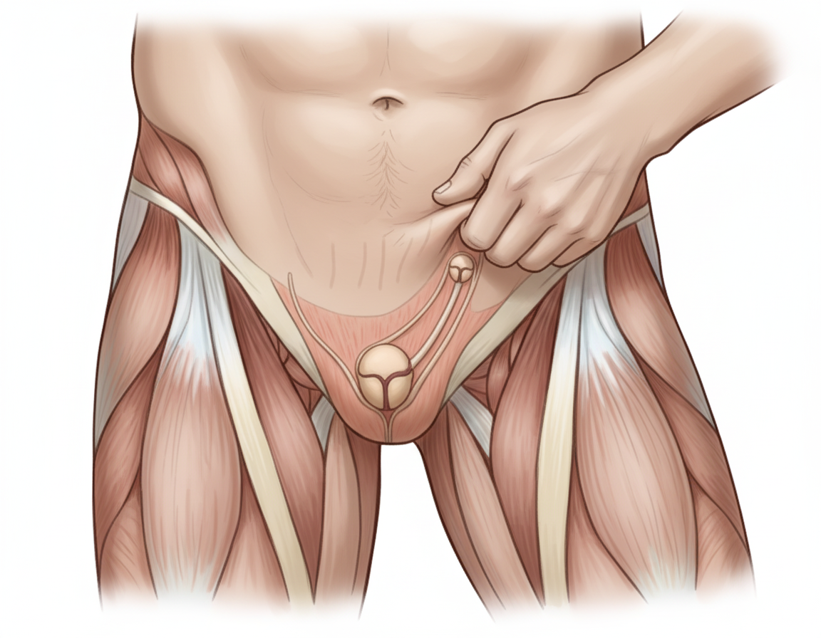
The image shows performance of the Cremasteric Reflex with ipsilateral elevation of testis. Which nerve mediates this reflex? Additional information: - Afferent: Femoral branch of genitofemoral nerve - Efferent: Genital branch of genitofemoral nerve - The reflex is elicited by lightly stroking the skin on the medial aspect of the superior part of the thigh - Rapid elevation of the testis on the same side confirms the cremasteric reflex - This reflex is extremely active in children

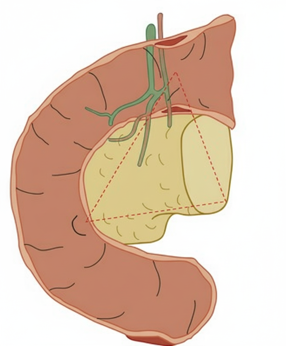
The image shows a triangular anatomical region with the following boundaries: 1. Confluence of cystic duct with common hepatic duct 2. Junction of head and body of pancreas 3. Junction of second and third parts of duodenum. Identify the structure:

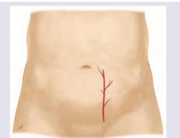
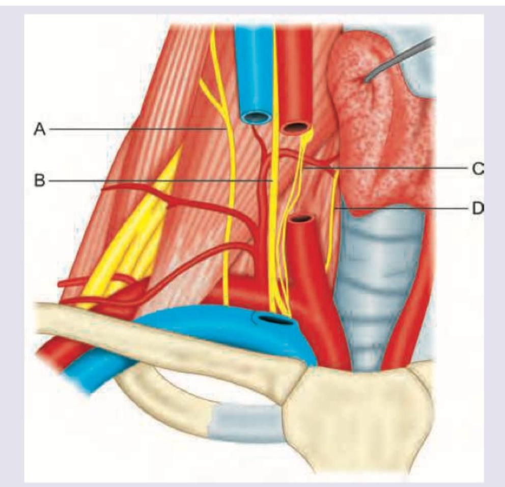
Which artery shown here should be avoided during paracentesis?

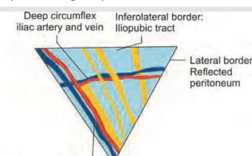
Which of the nerves shown in yellow color is not a part of the triangle of pain?

In this diagram, identify the structure whose paralysis causes decrease in respiratory movements: (AIIMS May 2016)

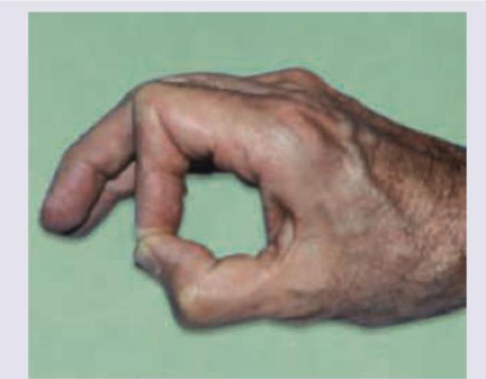
Identify the nerve damaged in the picture:

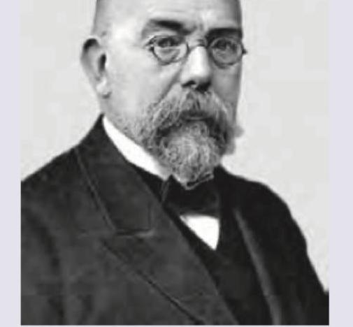
Identify the scientist. (Recent NEET Pattern 2016-17)

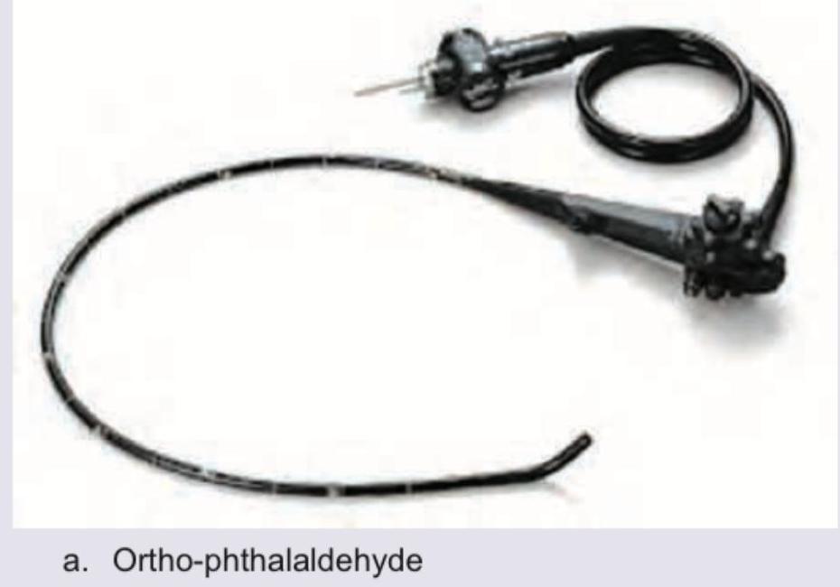
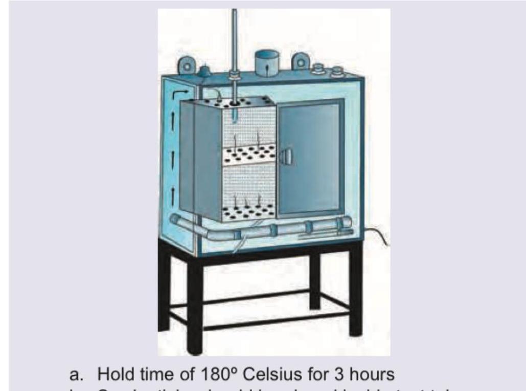
The following instrument is sterilized using:

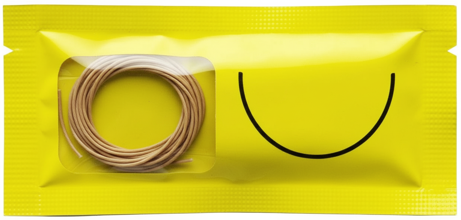
The suture material shown below is sterilized by which method?

All are correct about the method of sterilization shown here except:

Explanation: ***Genitofemoral nerve*** - The **cremasteric reflex** involves both the afferent (sensory) and efferent (motor) limbs mediated by branches of the **genitofemoral nerve**. - The afferent limb is the **femoral branch of the genitofemoral nerve**, which senses the stimulation on the inner thigh, and the efferent limb is the **genital branch of the genitofemoral nerve**, which causes the cremaster muscle to contract.
Explanation: ***Correct: Gastrinoma triangle (Passaro's triangle)*** - The **Gastrinoma triangle** or **Passaro's triangle** is a triangular region in the abdomen where the majority of gastrinomas (tumors that secrete gastrin) are found - Its boundaries are: - **Superiorly**: Confluence of the cystic duct with the common hepatic duct - **Inferiorly**: Junction of the head and body of the pancreas - **Medially**: Junction of the second and third parts of the duodenum - This is the most common location for **gastrinomas** in Zollinger-Ellison syndrome - Approximately **90% of gastrinomas** are found within this triangle *Incorrect: Calot's triangle* - Also known as the **hepatocystic triangle** - Boundaries: Cystic duct, common hepatic duct, and inferior surface of the liver - Important surgical landmark during **cholecystectomy** for identifying the cystic artery *Incorrect: Hesselbach's triangle* - Also known as the **inguinal triangle** - Boundaries: Inferior epigastric vessels laterally, rectus abdominis medially, and inguinal ligament inferiorly - Site of **direct inguinal hernias** *Incorrect: Triangle of Petit* - Also known as the **inferior lumbar triangle** - Boundaries: Iliac crest inferiorly, latissimus dorsi medially, and external oblique laterally - Potential site of **lumbar hernias**
Explanation: ***Inferior epigastric artery*** - The image depicts the **inferior epigastric artery** and its branches, which run on the anterior abdominal wall. These vessels are susceptible to injury during paracentesis, particularly if the needle is inserted lateral to the rectus abdominis muscle. - Injury to the inferior epigastric artery can lead to **significant bleeding** and **hematoma formation**, even after careful technique. *Superior epigastric artery* - The **superior epigastric artery** is a continuation of the internal thoracic artery and supplies the upper rectus abdominis muscle. It is located more superiorly and less commonly injured during **paracentesis** which is typically performed in the lower abdomen. - While it anastomoses with the inferior epigastric artery, its anatomical position makes it a less likely candidate for direct injury during standard paracentesis procedures which are usually performed below the umbilicus. *Superior hypogastric artery* - The **superior hypogastric artery** (also known as the median sacral artery) originates from the posterior aspect of the aorta and is involved in the arterial supply of structures within the pelvis. - This artery is located **deep within the pelvis**, far from the anterior abdominal wall where paracentesis is performed, making it anatomically irrelevant to this procedure. *Inferior hypogastric artery* - The term **inferior hypogastric artery** is not a standard anatomical term for a major artery; the primary pelvic arterial supply is from the internal iliac artery, sometimes referred to as the hypogastric artery, which branches into numerous smaller vessels. - Regardless of specific nomenclature, any such vessel would be located **deep in the pelvis** and not in the abdominal wall where injury during paracentesis would be a concern.
Explanation: ***Femoral nerve*** The **femoral nerve** is located laterally to the **femoral artery** and typically runs within the **iliopsoas groove**, outside the boundaries of the **triangle of pain**. Its position is more posterior and lateral, making it less vulnerable to injury during inguinal hernia repair compared to the nerves that traverse the "triangle of pain". *Lateral femoral cutaneous nerve* The **lateral femoral cutaneous nerve** is consistently found within the boundaries of the **triangle of pain**, increasing its susceptibility to injury during Lichtenstein hernia repair. Damage to this nerve can lead to **meralgia paraesthetica**, characterized by burning pain and numbness in the lateral thigh. *Femoral branch of Genitofemoral nerve* The **femoral branch of the genitofemoral nerve** typically crosses the **deep inguinal ring** and lies within the lateral part of the **triangle of pain**. Injury to this nerve during hernia repair can result in numbness or altered sensation in the anterior thigh. *Genital branch of Genitofemoral nerve* The **genital branch of the genitofemoral nerve** is located within the medial and inferior aspects of the **triangle of pain**. Damage to this nerve most commonly causes numbness or pain in the scrotum/labia majora and medial thigh.
Explanation: ***C*** - Structure C points to the **phrenic nerve** (C3-C5: "C3, 4, 5 keeps the diaphragm alive"), which primarily innervates the **diaphragm**. - The diaphragm is the **most important muscle of respiration**, responsible for approximately **70-80% of the work of breathing** during quiet respiration. - Paralysis of the phrenic nerve leads to **diaphragmatic paralysis**, significantly decreasing respiratory movements and causing respiratory compromise. *A* - Structure A points to a nerve arising from the **carotid sheath region**, likely representing a branch of the **cervical plexus** or a sympathetic nerve. - While these nerves have various functions (e.g., cutaneous innervation, vasomotor control), their paralysis does not directly cause a significant decrease in overall respiratory movements. *B* - Structure B points to the **subclavian artery**, which is a major blood vessel supplying blood to the upper limb and head/neck region. - Paralysis is a neurological term and does not apply to an artery; therefore, its occlusion or damage would lead to **ischemia** of supplied tissues, not a direct decrease in respiratory movements. *D* - Structure D points to the **trachea**, which is the main airway leading to the lungs. - The trachea is a cartilaginous tube, not a nerve or muscle that can be paralyzed; its obstruction would lead to **dyspnea** and respiratory distress, not decreased movements due to paralysis.
Explanation: ***Median*** - The image illustrates a patient unable to form a perfect "O" shape with their thumb and index finger, a sign known as the **"A-OK" sign** or **"pinch sign"**, indicating weakness of the **flexor pollicis longus** and **flexor digitorum profundus** (index finger) muscles. - These muscles are innervated by the **anterior interosseous nerve (AIN)**, which is a pure motor branch of the **median nerve**. *Radial* - Radial nerve damage typically results in a **wrist drop** due to paralysis of the wrist extensors and finger extensors. - The patient in the image is holding their wrist and fingers in flexion, which is not characteristic of radial nerve palsy. *Ulnar* - Ulnar nerve damage would result in a **"claw hand" deformity**, where the 4th and 5th fingers are hyperextended at the metacarpophalangeal joints and flexed at the interphalangeal joints. - It would also affect the **adduction of the thumb** (Froment's sign) and the intrinsic muscles of the hand, which are not the primary issues depicted. *Posterior interosseous nerve* - Damage to the posterior interosseous nerve (PIN), a branch of the radial nerve, primarily affects the **extensors of the fingers and thumb** leading to an inability to extend them. - It does not typically affect the **flexor pollicis longus** or **flexor digitorum profundus to the index finger**, which are responsible for the 'pinch' movement shown.
Explanation: ***Robert Koch*** - The image provided is a portrait of **Robert Koch**, a German physician and microbiologist. - He is known for his work on identifying the **causative agents of diseases** such as tuberculosis, cholera, and anthrax. *Louis Pasteur* - **Louis Pasteur** was a French chemist and microbiologist, known for his discoveries of the principles of **vaccination, microbial fermentation, and pasteurization**. - His appearance, particularly his facial features and style of beard, is distinct from the person in the image. *Christiaan Barnard* - **Christiaan Barnard** was a South African cardiac surgeon who performed the **world's first successful human-to-human heart transplant**. - He lived much later than the person pictured and has a distinct appearance. *William Osler* - **William Osler** was a Canadian physician and one of the four founding professors of **Johns Hopkins Hospital**. He is often called the "Father of Modern Medicine". - While a prominent figure in medicine, his physical appearance in historical photographs is different from the individual shown.
Explanation: ***Ortho-phthalaldehyde (OPA)*** - The instrument shown is an **endoscope**, which is a heat-sensitive, semi-critical device requiring high-level disinfection. - **OPA** is a common and effective high-level disinfectant used for endoscopes, as it offers a rapid disinfection time, broad microbicidal activity, and is stable. *Ethylene dioxide* - **Ethylene oxide (EO)** is a gas sterilant primarily used for heat and moisture-sensitive medical devices that cannot be effectively disinfected by liquid chemicals. - EO sterilization requires specific, highly controlled chambers and aeration periods to remove toxic residuals, making it less practical for routine endoscope disinfection in clinical settings compared to liquid agents. *Beta-propiolactone* - **Beta-propiolactone** is a chemical sterilant with broad-spectrum activity but is notable for being a probable human carcinogen and a strong irritant. - Due to its significant toxicity and potential health risks, its use in medical sterilization, particularly for instruments like endoscopes, has largely been phased out or is very restricted. *Formaldehyde* - **Formaldehyde** is a strong disinfectant and sterilant that can be used in both liquid and gaseous forms; however, it is a known carcinogen and has strong irritating fumes. - While effective, its high toxicity and safety concerns limit its routine use for endoscopes, especially when less hazardous high-level disinfectants like OPA are available.
Explanation: ***Gamma radiation*** - **Catgut sutures**, being made from natural, collagen-based materials, are **heat-sensitive** and cannot withstand high temperatures or steam. - **Ionizing radiation**, specifically gamma radiation, is effective for sterilizing heat-sensitive medical devices and materials without causing thermal damage, while still effectively killing microorganisms. *Dry heat sterilization* - Dry heat sterilization requires high temperatures for extended periods, which would **damage the protein structure of catgut**, leading to degradation and loss of tensile strength. - This method is typically used for items that can withstand high heat but are sensitive to moisture, such as powders, oils, and some glassware. *Autoclave (moist heat)* - While highly effective for many surgical instruments, **autoclaving (moist heat)** would cause the **collagen fibers of catgut to denature** and melt, destroying the suture material. - Catgut is highly susceptible to hydrolysis and degradation in the presence of heat and moisture. *Chemical sterilization with ethylene oxide* - Although ethylene oxide can be used for heat-sensitive materials, its use is often limited due to **toxic residue concerns** and the need for a lengthy aeration process. - While technically possible for some heat-sensitive items, **gamma radiation** is generally preferred for catgut due to its efficiency and lack of toxic residues.
Explanation: ***Biological indicator is Bacillus subtilis strips containing spores*** - This statement is **INCORRECT** for hot air oven (dry heat) sterilization, making it the correct answer to this EXCEPT question. - The appropriate biological indicator for dry heat sterilization is **Bacillus atrophaeus** (formerly *Bacillus subtilis var. niger*), which has spores with high heat resistance. - *Bacillus subtilis* strips are used as biological indicators for **ethylene oxide sterilization**, not dry heat. *Hold time of 180° Celsius for 3 hours* - While this temperature-time combination is **excessive**, it would still achieve sterilization (though inefficient). - Standard protocols: **180°C for 30 minutes**, 170°C for 1 hour, or 160°C for 2 hours. - The extended time doesn't make the statement fundamentally incorrect for sterilization purposes. *Swab sticks should be placed inside test tubes* - This statement is **CORRECT** for dry heat sterilization. - Swab sticks are placed in test tubes with cotton plugs or loose caps to allow heat penetration while preventing recontamination after sterilization. *Chemical indicator of sterilization is Browne's tube number 3* - While **Browne's tube No. 1** is more specific for hot air ovens, Browne's tube No. 3 can indicate exposure to high temperatures. - However, Browne's tube No. 3 is specifically designed for **steam sterilization at 121°C**, making this statement technically imprecise but not the primary exception. - The biological indicator error is more fundamentally incorrect.
Anatomical Basis of Common Clinical Conditions
Practice Questions
Surgical Anatomy
Practice Questions
Anatomical Basis of Trauma
Practice Questions
Anatomical Aspects of Infections
Practice Questions
Anatomical Considerations in Regional Anesthesia
Practice Questions
Anatomical Basis of Physical Examination
Practice Questions
Clinical Correlations in Neuroanatomy
Practice Questions
Anatomical Approaches in Minimally Invasive Procedures
Practice Questions
Imaging Correlations in Clinical Anatomy
Practice Questions
Anatomical Variations of Clinical Importance
Practice Questions
Get full access to all questions, explanations, and performance tracking.
Start For Free