Garlicky odor in the gastric contents is characteristic of which non-metallic poisoning?
A patient presented to OPD with ophthalmoplegia and ptosis. Diagnosis of superior orbital fissure syndrome was confirmed after examination. Which nerves are compressed in this case ?
Which of the following veins is involved in the formation of esophageal varices?
A woman presents with a breast lump, associated with skin dimpling and nipple retraction. What is the most likely anatomical structure responsible for the skin dimpling?
A patient presents with loss of sensation on the posterior surface of the ear along with a lesion. Which structure is most likely involved?
A 55-year-old man is diagnosed with left testicular carcinoma. Which of the following lymph nodes is the first to be involved?
A patient presented to the OPD with an abnormal gait. On examination, a right-sided hip drop was observed when the patient was asked to stand on the left foot. Which of the following muscles are paralyzed in this patient?
A middle-aged lady choked while eating fish and has associated symptoms of coughing, hoarseness of voice, and a foreign body sensation in the throat. On examination, the pyriform fossa is found to be inflamed. Which of the following nerves supplies this region?
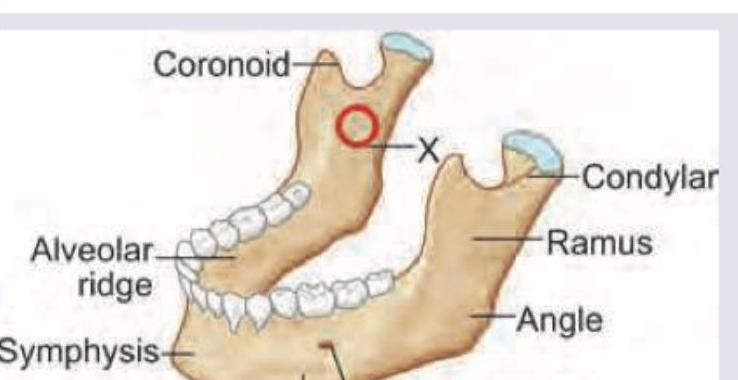
While giving inferior alveolar block, the injection is given above which foramen marked as X ?

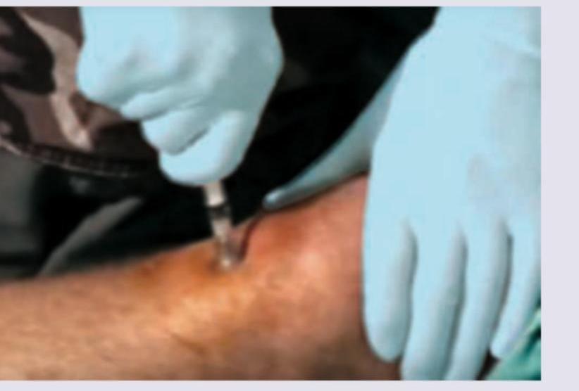
All of the following sites are used for the route of adrenaline administration shown below except:

Explanation: **Explanation:** The characteristic **garlicky odor** in gastric contents or breath is a classic clinical sign of **Phosphorus poisoning** (specifically Yellow Phosphorus). Yellow phosphorus is a highly toxic substance often found in rodenticides and fireworks. When ingested, it causes severe gastrointestinal irritation and hepatotoxicity. The odor is a result of the volatile phosphine gas produced during the breakdown of phosphorus compounds in the body. **Analysis of Options:** * **Phosphorus (Correct):** It is a non-metallic element known for its distinct garlic-like smell. In forensic medicine, this is a high-yield diagnostic feature during gastric lavage or autopsy. * **Sulphur:** Typically associated with the smell of **rotten eggs** (due to Hydrogen Sulphide gas), not garlic. * **Iodine:** Ingestion usually results in a characteristic **metallic taste** and stains the mucous membranes brown/blue; it does not produce a garlic odor. * **Chlorine:** Known for its pungent, **bleaching, or swimming pool-like** suffocating odor. **Clinical Pearls for NEET-PG:** * **Garlicky Odor Differentiator:** While Phosphorus is a non-metal, **Arsenic** (a metalloid) and **Organophosphates** (insecticides) also produce a garlicky odor. Always check if the question specifies "non-metallic." * **Luminous Vomitus:** Gastric contents in phosphorus poisoning may show **phosphorescence** (glow in the dark), a unique diagnostic clue. * **Hepatotoxicity:** Phosphorus poisoning typically leads to "acute yellow atrophy" of the liver and fulminant hepatic failure. * **Other Odors to Remember:** * **Bitter Almonds:** Cyanide * **Rotten Eggs:** Hydrogen Sulphide * **Kerosene-like:** Organophosphates (due to the solvent) * **Fruity:** Ethanol/Ketoacidosis
Explanation: ***III, IV, V1, VI*** - The **superior orbital fissure** is a critical anatomical passage that transmits the **oculomotor nerve (III)**, **trochlear nerve (IV)**, the **ophthalmic division of the trigeminal nerve (V1)**, and the **abducens nerve (VI)**. - Compression of these nerves collectively results in **ophthalmoplegia** (paralysis of eye muscles due to III, IV, VI involvement [1]) and **ptois** (drooping of the upper eyelid due to III involvement), which are the classic signs of superior orbital fissure syndrome. *III, IV, V1, V2* - This option is incorrect because the **maxillary division of the trigeminal nerve (V2)** does not pass through the superior orbital fissure. - V2 exits the skull through the **foramen rotundum** to supply the maxillary region, and is therefore not affected in this syndrome. *II, III, IV, VI* - This option is incorrect because the **optic nerve (II)** is not involved in superior orbital fissure syndrome. - The optic nerve passes through the **optic canal**, a separate opening. Involvement of the optic nerve would cause vision loss and indicate a more extensive condition like **orbital apex syndrome**. *II, III, IV, V1* - This is incorrect as it includes the **optic nerve (II)**, which, as mentioned, travels through the optic canal, not the superior orbital fissure. - The absence of vision loss or an **afferent pupillary defect** helps differentiate superior orbital fissure syndrome from pathologies involving the optic nerve.
Explanation: ***Left gastric vein*** - The **left gastric vein** (coronary vein) is the primary vessel involved in esophageal varices formation through **portosystemic anastomoses** at the **gastroesophageal junction** during portal hypertension [1]. - It connects the **portal circulation** to the **systemic circulation** via esophageal veins, creating the most clinically significant pathway for variceal development [1]. *Left gastroepiploic vein* - This vein drains the **greater curvature of the stomach** and flows into the splenic vein, not forming significant connections with esophageal circulation. - It does not participate in **portosystemic anastomoses** at the esophageal level where varices typically develop. *Right gastric vein* - Drains the **lesser curvature of the stomach** and flows directly into the portal vein [2], with minimal anatomical connection to esophageal vessels. - Does not form the critical **portosystemic anastomoses** necessary for esophageal varices formation during portal hypertension. *Right gastroepiploic vein* - Drains the **greater curvature of the stomach** and connects to the superior mesenteric vein, distant from esophageal circulation. - Lacks the anatomical connections required for **portosystemic anastomoses** at the gastroesophageal junction where varices develop.
Explanation: ***Suspensory (Cooper’s) ligaments*** - These are fibrous septa that run from the deep pectoral fascia to the dermis of the skin, providing structural support to the breast [1]. - Invasion and shortening of these ligaments by a growing tumor pull on the overlying skin, causing the characteristic **skin dimpling** or peau d'orange appearance [1]. *Lactiferous ducts* - These are the milk ducts that converge and open at the nipple [1]. - Malignant infiltration of the lactiferous ducts is more commonly associated with **nipple retraction** and pathologic nipple discharge, rather than skin dimpling [2]. *Pectoral fascia* - This is a deep layer of connective tissue that covers the pectoralis major muscle, on which the breast lies [1]. - Tumor invasion into the pectoral fascia can cause the breast to become **fixed** to the chest wall, a sign of advanced disease, but does not directly cause superficial skin dimpling. *Subcutaneous fat* - This tissue makes up the bulk of the breast volume and surrounds the glandular components. - Subcutaneous fat itself lacks the tensile strength to pull the skin inward; it is the **fibrous ligaments** passing through the fat that cause retraction [1].
Explanation: ***Greater Auricular Nerve***- The **Greater Auricular Nerve (GAN)**, originating from the cervical plexus (C2, C3), provides sensory innervation to the **skin over the mastoid process** and the **posterior surface of the auricle (ear)**.- Damage (lesion involvement) to this nerve results specifically in **sensory loss (anesthesia)** in its distribution area, matching the patient's presentation.*Internal Jugular Vein*- This is a large deep vein responsible for major **venous drainage** of the head and neck, not sensory innervation.- Involvement would cause **venous congestion** or potentially severe complications related to thrombosis, not isolated sensory loss.*External Jugular Vein*- This is a superficial vein responsible for minor **venous drainage** of the face and head, running lateral to the sternocleidomastoid muscle.- Any compromise to this vein affects the circulatory system (**venous return**) and does not lead to sensory deficits.*External Carotid Artery*- This is a major artery supplying the extracranial structures of the head; its primary function is **blood supply (perfusion)**.- Lesions would typically cause signs of **ischemia** or hemorrhage in its distribution, not an isolated nerve-related sensory loss on the posterior ear.
Explanation: ***Para-aortic lymph nodes***- The testes originate in the retroperitoneum, and their lymphatic drainage follows the **gonadal vessels**, primarily draining to the **paraaortic** (L1/L2 level) nodes, irrespective of descent into the scrotum.- For the **left testis**, the primary landing site for metastatic cancer is the **para-aortic chain** due to its drainage pathway along the left gonadal vein into the left renal vein [1].*Inguinal lymph nodes*- These nodes drain the **scrotal skin**, coverings, and structures superficial to the **tunica vaginalis**, not the testis itself [1].- Involvement of inguinal nodes only occurs late in the disease if the tumor has invaded the scrotal wall, or post-scrotal trauma/surgery [1].*Iliac lymph nodes*- These nodes (internal and external) primarily drain the **pelvic structures** (e.g., bladder, prostate, lower limb).- They are considered second-echelon nodes for testicular cancer, typically involved only after spread to the primary retroperitoneal (para-aortic/paracaval) chains.*Pre-caval lymph nodes*- These nodes are the primary landing site for the **right testicular carcinoma** because the right testicular vein drains directly into the **Inferior Vena Cava (IVC)**.- For the left testis, the drainage is primarily routed to the **para-aortic** nodes, although some crossover to the pre-caval nodes may occur later.
Explanation: ***Left gluteus medius and gluteus minimus*** - The Trendelenburg sign is positive when the pelvis drops on the side opposite the one being stood on (the unsupported side). - This indicates paralysis or weakness of the **hip abductors** on the **standing leg** (left side in this case), which are the gluteus medius and gluteus minimus, responsible for stabilizing the pelvis. - These muscles are innervated by the **superior gluteal nerve**. *Right gluteus maximus and gluteus medius* - Paralysis of muscles on the right side would typically manifest as a deficit when bearing weight on the **right leg** (causing the left hip to drop). - The **gluteus maximus** is mainly a powerful hip extensor (needed for climbing stairs, running, and rising from a seated position) and does not play the primary role in stabilizing the pelvis during single-leg stance. *Right gluteus medius and gluteus minimus* - If these muscles on the right side were paralyzed, the pelvis would drop on the **left side** when the patient attempts to stand on the right foot. - The finding of a right hip drop while standing on the left foot confirms the deficit is on the ipsilateral side of the standing limb (the **left side**). *Left gluteus maximus and gluteus medius* - While the **left gluteus medius** is correctly identified as being paralyzed, the inclusion of the gluteus maximus is inaccurate. - The **gluteus maximus** is innervated by the **inferior gluteal nerve** (not the superior gluteal nerve), and its paralysis causes difficulty with activities requiring hip extension, such as climbing stairs or rising from a chair (gluteus maximus lurch). - The Trendelenburg sign specifically tests the **superior gluteal nerve** and the hip abductors (gluteus medius and minimus), not the gluteus maximus.
Explanation: ***Internal Laryngeal Nerve (ILN)*** - The **Internal Laryngeal Nerve** (sensory branch of **superior laryngeal nerve**, CN X) provides **sensory innervation to the pyriform fossa** and surrounding hypopharyngeal structures. - Fish bone impaction in the pyriform fossa irritates the **ILN**, causing **cough reflex**, **hoarseness**, and **foreign body sensation** as described in this case. *External Laryngeal Nerve (ELN)* - The **External Laryngeal Nerve** is primarily a **motor nerve** that supplies the **cricothyroid muscle** for vocal cord tensioning. - It provides **minimal sensory contribution** and does not innervate the mucosa of the pyriform fossa. *Glossopharyngeal Nerve* - The **Glossopharyngeal Nerve (CN IX)** provides sensory innervation to the **oropharynx** and **posterior third of tongue**, not the pyriform fossa. - The pyriform fossa is anatomically part of the **hypopharynx (laryngopharynx)**, which is supplied by branches of the **vagus nerve**, not CN IX. *Recurrent Laryngeal Nerve* - The **Recurrent Laryngeal Nerve** primarily provides **motor innervation** to intrinsic laryngeal muscles (except cricothyroid). - Its sensory distribution is limited to the **infraglottic larynx** (below vocal cords), not the pyriform fossa region.
Explanation: ***Mandibular foramen*** - The image shows the **medial aspect of the mandibular ramus**, and the structure labeled 'X' points directly to the **mandibular foramen**, an opening on the internal surface of the ramus. - The **inferior alveolar nerve** enters the mandible through the mandibular foramen, making this the target for an **inferior alveolar nerve block** to anesthetize the mandibular teeth. *Mental foramen* - The **mental foramen** is located on the **buccal (outer) surface of the mandible**, typically between the first and second premolars, much further anteriorly than indicated by 'X'. - An injection near the mental foramen provides anesthesia for the **buccal soft tissues** and some anterior teeth, but not a widespread inferior alveolar block. *Inferior orbital foramen* - The **inferior orbital foramen** is located on the floor of the orbit, under the eye, and is completely unrelated to the mandible. - This foramen transmits the **infraorbital nerve**, which supplies sensation to the lower eyelid, side of the nose, and upper lip. *Incisive foramen* - The **incisive foramen** is located on the palate, posterior to the central incisors, and is part of the maxilla, not the mandible. - This foramen transmits the **nasopalatine nerve**, which supplies sensation to the anterior palatal mucosa.
Explanation: ***Ischial spine*** - The ischial spine is a pelvic landmark and is absolutely **not a site for intraosseous (IO) access** due to its deep location, proximity to vital structures, and lack of accessible bone marrow. - Using the ischial spine for IO access would be **dangerous and ineffective**, risking injury to nerves, blood vessels, and adjacent organs. *2 cm below tibial tuberosity* - This is a common and **appropriate site for intraosseous (IO) access** in adults and children. - The **proximal tibia** offers readily accessible bone marrow and a strong cortical bone for needle insertion. *2 cm superior to medial malleolus* - This location on the **distal tibia** is another recognized and suitable site for IO access, particularly in pediatric patients or when other sites are unavailable. - It provides a safe and effective entry point into the **bone marrow cavity**. *Greater tubercle of humerus* - The **proximal humerus**, near the greater tubercle, is a crucial and increasingly preferred site for IO access, especially in adults. - It offers a rapid flow rate and is often chosen in **emergency situations** when IV access is difficult to obtain.
Anatomical Basis of Common Clinical Conditions
Practice Questions
Surgical Anatomy
Practice Questions
Anatomical Basis of Trauma
Practice Questions
Anatomical Aspects of Infections
Practice Questions
Anatomical Considerations in Regional Anesthesia
Practice Questions
Anatomical Basis of Physical Examination
Practice Questions
Clinical Correlations in Neuroanatomy
Practice Questions
Anatomical Approaches in Minimally Invasive Procedures
Practice Questions
Imaging Correlations in Clinical Anatomy
Practice Questions
Anatomical Variations of Clinical Importance
Practice Questions
Get full access to all questions, explanations, and performance tracking.
Start For Free