Which of the following is NOT a composite muscle?
Pain of ovarian carcinoma is referred to which region?
The regionally named layer of tissue which encloses and binds muscle groups together is the:
After graft repair of a thoraco-abdominal aneurysm, a patient is unable to move both lower limbs. What is the most probable cause?
Atrophy of intrinsic muscles of the hand, sensory deficit on the medial side of the forearm and hand, and diminished radial pulse on turning the head to the affected side could be due to which of the following conditions?
Which maneuver is used to test for integrity of the long thoracic nerve?
Beevor's sign is typically associated with which group of muscles?
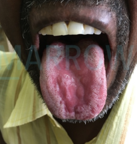
A 24-year-old male presents for a routine checkup. Radiographs reveal supernumerary teeth. General examination shows hypermobility of the shoulders, and the Gorlin sign is absent. What is the most probable diagnosis?
Which of the following is NOT a spiral muscle?
The condition shown in the color plate can be seen in which of the following syndromes?

Explanation: ### Explanation A **composite (or hybrid) muscle** is defined as a muscle that is supplied by more than one nerve, usually because it develops from more than one embryonic origin or spans across different functional compartments. **Why Gluteus Medius is the Correct Answer:** The **Gluteus medius** is not a composite muscle. It is supplied solely by the **superior gluteal nerve (L4, L5, S1)**. It acts primarily as an abductor and medial rotator of the hip and is crucial for stabilizing the pelvis during the swing phase of walking. **Why the other options are incorrect:** * **Brachialis:** It is a hybrid muscle supplied by the **Musculocutaneous nerve** (motor) and the **Radial nerve** (proprioceptive/sensory component to its lateral part). * **Adductor Magnus:** This is a classic composite muscle. Its adductor part is supplied by the **Obturator nerve**, while its "hamstring" part is supplied by the **Tibial component of the Sciatic nerve**. * **Flexor Digitorum Profundus (FDP):** It has a dual nerve supply. The medial half (digits 4 and 5) is supplied by the **Ulnar nerve**, and the lateral half (digits 2 and 3) is supplied by the **Anterior Interosseous branch of the Median nerve**. **High-Yield Clinical Pearls for NEET-PG:** * **Pectineus:** Often considered a hybrid muscle, supplied by the Femoral nerve and occasionally the Obturator nerve. * **Biceps Femoris:** Another hybrid; the long head is supplied by the Tibial part of the Sciatic nerve, while the short head is supplied by the Common Peroneal part. * **Digastric Muscle:** The anterior belly is supplied by the Nerve to Mylohyoid (CN V3), and the posterior belly by the Facial nerve (CN VII). * **Trendelenburg Sign:** Clinical correlation for Gluteus medius; paralysis leads to the dropping of the pelvis on the unsupported side.
Explanation: **Explanation:** The correct answer is **D. Medial surface of thigh.** **Underlying Medical Concept:** The ovary is embryologically derived from the same level as the kidneys and descends into the pelvis. However, its sensory innervation is closely related to the **obturator nerve (L2-L4)**. The ovary lies in the **ovarian fossa** on the lateral pelvic wall [1], and the obturator nerve runs directly lateral to this fossa, separated only by the parietal peritoneum. In cases of ovarian carcinoma or inflammation, the tumor or inflammatory exudate can irritate the obturator nerve. Since the obturator nerve provides sensory innervation to the **medial surface of the thigh** (via its cutaneous branch), the brain perceives the pain as originating from that skin area [3]. This is a classic example of **referred pain**. **Analysis of Incorrect Options:** * **A & B (Back of thigh and Gluteal region):** These areas are primarily supplied by the **posterior cutaneous nerve of the thigh** and branches of the **sacral plexus (S1-S3)**. These nerves are not in direct anatomical proximity to the ovarian fossa. * **C (Anterior surface of thigh):** This region is supplied by the **femoral nerve (L2-L4)**. While it shares the same spinal segments as the obturator nerve, the femoral nerve does not run in the ovarian fossa and is protected by the psoas major muscle [2]. **High-Yield Clinical Pearls for NEET-PG:** * **Howship-Romberg Sign:** Pain/paresthesia in the medial thigh due to obturator nerve compression (often seen in obturator hernias). * **Nerve at Risk:** The obturator nerve is the most commonly injured nerve during radical pelvic surgeries (like lymphadenectomy for ovarian cancer). * **Lymphatic Drainage:** Ovarian cancer primarily drains to the **Para-aortic lymph nodes** (not inguinal), following the ovarian arteries.
Explanation: **Explanation:** The correct answer is **Deep fascia**. **Why Deep Fascia is correct:** Deep fascia is a dense, organized connective tissue layer, devoid of fat, that lies deep to the skin and subcutaneous tissue. Its primary anatomical function is to invest (enwrap) muscles and bind them into functional groups. It is "regionally named" based on its location—for example, the *fascia lata* in the thigh or the *crural fascia* in the leg. By tightly enclosing muscles, it limits their outward expansion during contraction, which aids in the "musculovenous pump" mechanism to return blood to the heart. **Why the other options are incorrect:** * **Intermuscular septum:** While these are also made of dense connective tissue, they are inward extensions of the deep fascia that attach to bone. They separate muscle groups into compartments rather than enclosing the entire group as a whole. * **Neurovascular bundle:** This refers to a group of nerves and blood vessels (arteries and veins) that travel together through a body region, usually wrapped in a common connective tissue sheath. It does not bind muscle groups. * **Skin:** This is the outermost protective integument consisting of the epidermis and dermis. It covers the entire body but does not serve the specific mechanical function of binding muscle groups together. **High-Yield Clinical Pearls for NEET-PG:** * **Compartment Syndrome:** Because deep fascia is inelastic, excessive inflammation or bleeding within a muscle compartment increases pressure, potentially compressing nerves and vessels. This is a surgical emergency requiring a **fasciotomy**. * **Retinacula:** These are thickened bands of deep fascia near joints (like the wrist or ankle) that hold tendons in place, preventing "bowstringing" during movement. * **Investing Layer:** In the neck, the investing layer of deep cervical fascia encloses the trapezius and sternocleidomastoid muscles.
Explanation: **Explanation:** The clinical presentation of paraplegia following thoraco-abdominal aortic aneurysm (TAAA) repair is a classic complication caused by spinal cord ischemia [1]. **Why Option D is Correct:** The **Arteria Magna Radicularis (Artery of Adamkiewicz)** is the largest segmental medullary artery. It typically arises from a left-sided posterior intercostal artery between **T9 and L2**. It provides the primary blood supply to the lower two-thirds of the spinal cord by reinforcing the anterior spinal artery. During graft repair of a TAAA, this artery can be compromised due to clamping of the aorta or exclusion of the segmental vessels, leading to **Anterior Spinal Artery Syndrome**. This results in motor paralysis (loss of corticospinal tracts) and loss of pain/temperature sensation, while proprioception remains intact. **Why Incorrect Options are Wrong:** * **A. Thoracic splanchnic nerves:** These carry preganglionic sympathetic fibers to the abdomen. Damage would cause autonomic dysfunction (e.g., ileus), not motor paralysis. * **B. Lumbar spinal nerves:** While damage could cause weakness, it would typically result in lower motor neuron signs in specific dermatomes/myotomes. TAAA repair involves the cord blood supply at a higher level, affecting the entire distal cord. * **C. Ischemia of the lower limbs:** This would present with the "6 Ps" (Pain, Pallor, Pulselessness, etc.). While limbs would be difficult to move, the primary neurological deficit (paraplegia) in the context of aortic surgery is centrally mediated via the spinal cord [1]. **High-Yield Facts for NEET-PG:** * **Artery of Adamkiewicz:** Most common origin is **T9-T12** on the **left side**. * **Anterior Spinal Artery Syndrome:** Characterized by "Dissociated Sensory Loss" (loss of motor and pain/temp; preservation of vibration/position). * **Vulnerability:** The mid-thoracic spinal cord (T4-T8) is a "watershed area" and is most susceptible to global ischemia.
Explanation: **Explanation:** The clinical presentation described is a classic case of **Thoracic Outlet Syndrome (TOS)**, specifically caused by a **Cervical Rib**. **1. Why Cervical Rib is Correct:** A cervical rib is a supernumerary rib arising from the C7 vertebra. It can compress two vital structures passing through the scalene triangle: [1] * **Lower Trunk of the Brachial Plexus (C8-T1):** Compression leads to atrophy of the intrinsic muscles of the hand (T1) and sensory loss along the medial aspect of the forearm and hand (C8). [1] * **Subclavian Artery:** Compression results in vascular symptoms. The **Adson’s Test** (diminished radial pulse upon turning the head toward the affected side and taking a deep breath) is a hallmark clinical sign of this compression. [1] **2. Why Incorrect Options are Wrong:** * **Carpal Tunnel Syndrome:** Involves compression of the **median nerve** at the wrist. It causes wasting of the thenar eminence and sensory loss in the lateral 3.5 fingers, but does not affect the forearm or the radial pulse. * **Enlarged Axillary Lymph Nodes:** While these can compress parts of the brachial plexus, they typically affect the cords rather than the lower trunk specifically, and they rarely cause positional obliteration of the radial pulse. * **Supracondylar Fracture of Humerus:** Usually leads to injury of the median or radial nerve and may cause **Volkmann’s Ischemic Contracture** (brachial artery involvement), but it does not present with symptoms related to head movement. **Clinical Pearls for NEET-PG:** * **Adson’s Test:** Positive in Cervical Rib/TOS (radial pulse disappears). [1] * **Gilliatt-Sumner Hand:** The specific pattern of hand wasting seen in TOS. * **Nerve involved:** Primarily the **T1** root (Lower Trunk), leading to Klumpke’s-like palsy symptoms.
Explanation: **Explanation:** The **Long Thoracic Nerve** (C5, C6, C7) supplies the **Serratus Anterior** muscle. The primary functions of this muscle are protraction of the scapula and, crucially, **upward rotation of the glenoid cavity**. This upward rotation is essential for abducting the arm beyond 90 degrees. Therefore, asking a patient to **raise their arm above their head** (overhead abduction) is the definitive clinical test for the integrity of this nerve. **Analysis of Options:** * **Option A (Touching opposite shoulder):** This involves adduction and internal rotation, primarily testing the Pectoralis major and Subscapularis, not the Serratus anterior. * **Option B (Shoulder shrug):** This tests the **Trapezius** muscle, which is supplied by the **Spinal Accessory Nerve (CN XI)**. * **Option D (Lifting an object):** This primarily involves the Biceps brachii, Brachialis, and back extensors; it is not a specific test for the long thoracic nerve. **Clinical Pearls for NEET-PG:** * **Winging of Scapula:** Damage to the long thoracic nerve (often during radical mastectomy or chest tube insertion) causes the medial border of the scapula to protrude posteriorly, especially when the patient pushes against a wall. * **Mnemonic:** "C5, 6, 7 raise your arms to heaven" (refers to the nerve roots and the action of overhead abduction). * **Saltatorial Nerve:** The long thoracic nerve is also known as the "Nerve of Bell." * **Dual Action:** The Serratus anterior is often called the "Boxer’s muscle" because it is the main protractor of the scapula used during punching.
Explanation: Explanation: Beevor’s sign is a clinical finding characterized by the upward movement of the umbilicus when a patient attempts to flex the neck or perform a sit-up from a supine position. This occurs due to selective weakness of the lower abdominal muscles (supplied by T10–T12 spinal nerves) relative to the upper abdominal muscles (supplied by T7–T9). When the upper segments contract while the lower segments are paralyzed, the umbilicus is pulled superiorly toward the stronger muscles. * Why Option A is correct: The sign specifically tests the integrity of the rectus abdominis and the spinal levels T7 through T12. It is most classically associated with spinal cord lesions at the T10 level or in patients with Facio-Scapulo-Humeral Dystrophy (FSHD). * Why Options B, C, and D are incorrect: While facial (B) and hand (D) muscles are involved in various neurological signs (e.g., Chvostek’s or Froment’s sign), they do not influence umbilical movement. Respiratory muscles (C), like the diaphragm, are involved in breathing mechanics but do not cause the localized abdominal shift seen in Beevor’s sign. High-Yield Clinical Pearls for NEET-PG: 1. Localization: A positive Beevor’s sign suggests a lesion between T10 and T12. 2. Key Association: It is a highly specific clinical marker for Facio-Scapulo-Humeral Dystrophy (FSHD), often appearing early in the disease. 3. Differential Diagnosis: It can also be seen in Amyotrophic Lateral Sclerosis (ALS) and certain spinal cord tumors. 4. Inverted Beevor’s Sign: Downward movement of the umbilicus (rare) indicates upper abdominal muscle weakness.
Explanation: **Explanation:** The clinical presentation of **supernumerary teeth** and **hypermobility of the shoulders** (the ability to bring shoulders together anteriorly) is the classic hallmark of **Cleidocranial Dysplasia**, also known as **Marie-Sainton Disease**. **1. Why Marie-Sainton Disease is correct:** This is an autosomal dominant skeletal dysplasia caused by a mutation in the **RUNX2 gene**. Key features include: * **Clavicular Hypoplasia/Aplasia:** Leads to the characteristic hypermobility of shoulders. * **Dental Anomalies:** Delayed eruption of permanent teeth and multiple **supernumerary teeth**. * **Cranial Features:** Delayed closure of fontanelles and Wormian bones. * **Gorlin Sign:** While the "Gorlin sign" (touching the tip of the nose with the tongue) is often associated with Ehlers-Danlos, its absence here helps rule out EDS, pointing specifically to the skeletal pathology of Marie-Sainton. **2. Why other options are incorrect:** * **Sturge-Weber Syndrome:** A neurocutaneous disorder characterized by a Port-wine stain (Nevus Flammeus), leptomeningeal angiomas, and glaucoma. It does not involve clavicular or supernumerary tooth anomalies. * **Hallermann-Streiff Syndrome:** Characterized by "bird-like" facies, microphthalmia, congenital cataracts, and hypotrichosis. While dental anomalies occur, shoulder hypermobility is not a feature. * **Ehlers-Danlos Syndrome (EDS):** While EDS presents with joint hypermobility and a positive Gorlin sign (due to a long/flexible lingual frenulum), it does not typically feature supernumerary teeth or clavicular aplasia. **NEET-PG High-Yield Pearls:** * **RUNX2 Gene:** The specific molecular marker for Cleidocranial Dysplasia. * **Wormian Bones:** Frequently tested association with this condition. * **Shoulder Sign:** If a patient can touch their shoulders in the midline, think Marie-Sainton.
Explanation: The concept of **spiral muscles** refers to muscles whose fibers undergo a twist or rotation between their origin and insertion. This structural arrangement allows for a greater range of motion and increased power during contraction. **1. Why Sternocleidomastoid is the Correct Answer:** The **Sternocleidomastoid (SCM)** is a **parallel-fibered muscle**. Its fibers run straight from the manubrium sterni and clavicle to the mastoid process without any twisting or spiraling. While it acts as a primary rotator of the head, its anatomical fiber orientation is linear, not spiral. **2. Analysis of Incorrect Options (Spiral Muscles):** * **Supinator:** This is a classic example of a spiral muscle. It arises from the lateral epicondyle and supinator crest and wraps spirally around the upper part of the radius to insert on its lateral surface. * **Pectoralis Major:** This is a **cruciate (spiral) muscle**. The fibers of the lower (sternocostal) head twist behind the upper (clavicular) head to insert into the lateral lip of the bicipital groove, resulting in a 180-degree twist. * **Trapezius:** The lower fibers of the trapezius spiral upwards and laterally to reach the tubercle of the scapular spine, making it a spiral muscle. **3. NEET-PG High-Yield Pearls:** * **Other Spiral Muscles:** Latissimus dorsi (twists around the teres major) and Tibialis anterior. * **Functional Significance:** Spiraling allows a muscle to exert force in multiple planes and prevents the muscle from becoming "slack" during complex joint movements. * **Pectoralis Major Clinical:** The twisting of the pectoralis major forms the rounded **anterior axillary fold**. Loss of this fold is a clinical sign of pectoralis major rupture or congenital absence (Poland Syndrome).
Explanation: ***Both Melkersson-Rosenthal syndrome and Down syndrome*** - **Fissured tongue** (scrotal tongue/lingua plicata) is a characteristic feature of both **Melkersson-Rosenthal syndrome** and **Down syndrome**. - In **Melkersson-Rosenthal syndrome**, it forms part of the classic triad with **facial paralysis** and **granulomatous cheilitis**, while in **Down syndrome**, it occurs in approximately **80%** of patients alongside other oral manifestations. *Melkersson-Rosenthal syndrome* - While **fissured tongue** is indeed present in this syndrome, it occurs alongside **facial nerve palsy** and **granulomatous lip swelling**. - This option is incomplete as it doesn't acknowledge that **Down syndrome** also commonly presents with **fissured tongue**. *Down syndrome* - Although **fissured tongue** is highly prevalent in Down syndrome patients (up to **80%**), this option alone is insufficient. - It fails to recognize that **Melkersson-Rosenthal syndrome** also characteristically presents with **fissured tongue** as part of its diagnostic triad. *Pierre Robin syndrome* - This syndrome is characterized by **micrognathia**, **cleft palate**, and **glossoptosis** (posterior displacement of tongue). - **Fissured tongue** is **not** a recognized feature of **Pierre Robin syndrome**, making this option incorrect.
Anatomical Basis of Common Clinical Conditions
Practice Questions
Surgical Anatomy
Practice Questions
Anatomical Basis of Trauma
Practice Questions
Anatomical Aspects of Infections
Practice Questions
Anatomical Considerations in Regional Anesthesia
Practice Questions
Anatomical Basis of Physical Examination
Practice Questions
Clinical Correlations in Neuroanatomy
Practice Questions
Anatomical Approaches in Minimally Invasive Procedures
Practice Questions
Imaging Correlations in Clinical Anatomy
Practice Questions
Anatomical Variations of Clinical Importance
Practice Questions
Get full access to all questions, explanations, and performance tracking.
Start For Free