
Clinical Anatomy — MCQs

On this page
During nasogastric tube insertion, obstruction is encountered at 40cm due to ?
Which nerve is least likely to be affected by entrapment syndrome?
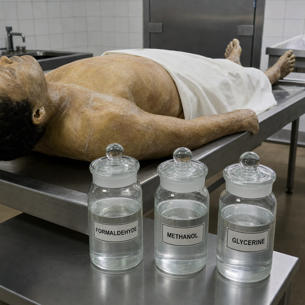
The procedure uses a fluid that contains a mixture of which of the following components?
All of the following are types of epiphysis, EXCEPT:
The regionally named layer of tissue which encloses and binds muscle groups together is the:
Practice by Chapter
Anatomical Basis of Common Clinical Conditions
Practice Questions
Surgical Anatomy
Practice Questions
Anatomical Basis of Trauma
Practice Questions
Anatomical Aspects of Infections
Practice Questions
Anatomical Considerations in Regional Anesthesia
Practice Questions
Anatomical Basis of Physical Examination
Practice Questions
Clinical Correlations in Neuroanatomy
Practice Questions
Anatomical Approaches in Minimally Invasive Procedures
Practice Questions
Imaging Correlations in Clinical Anatomy
Practice Questions
Anatomical Variations of Clinical Importance
Practice Questions
Want unlimited practice?
Get full access to all questions, explanations, and performance tracking.
Scan to download app