All (378)Anatomy (22)Anatomy (5)Anesthesiology (9)Behavioral Science (1)Biochemistry (3)Biochemistry (21)Biostatistics (4)Community Medicine (20)Dermatology (13)Diagnosis (3)ENT (11)Forensic Medicine (13)General Medicine (1)Internal Medicine (19)Internal Medicine (26)Microbiology (20)OB/GYN (16)Obstetrics and Gynecology (16)Ophthalmology (10)Orthopaedics (14)Pathology (25)Pediatrics (18)Pediatrics (2)Pharmacology (37)Physiology (10)Psychiatry (6)Radiology (12)Surgery (21)
Q261
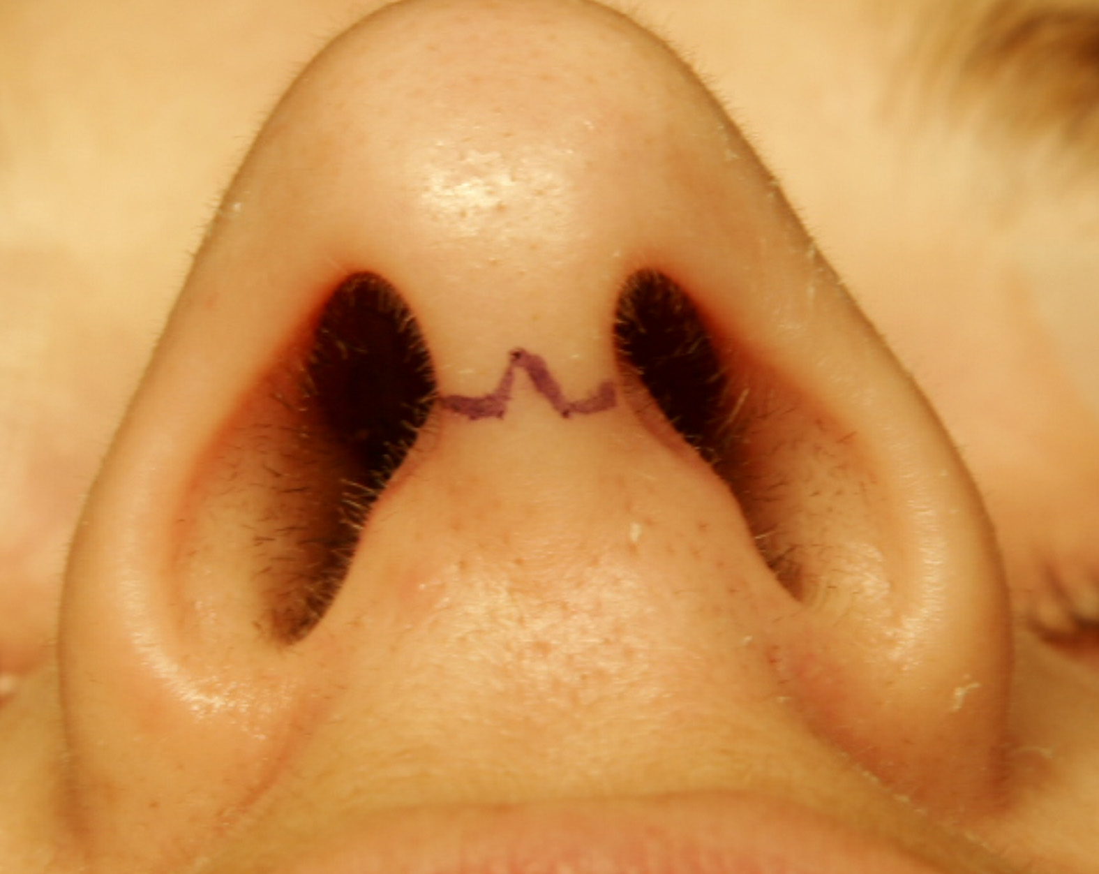
A nasal surgery was carried out with the incision shown in the image. What was the procedure likely carried out?
Q262
In a patient with right vestibular neuronitis, what will be the finding on the head impulse test?
Q263
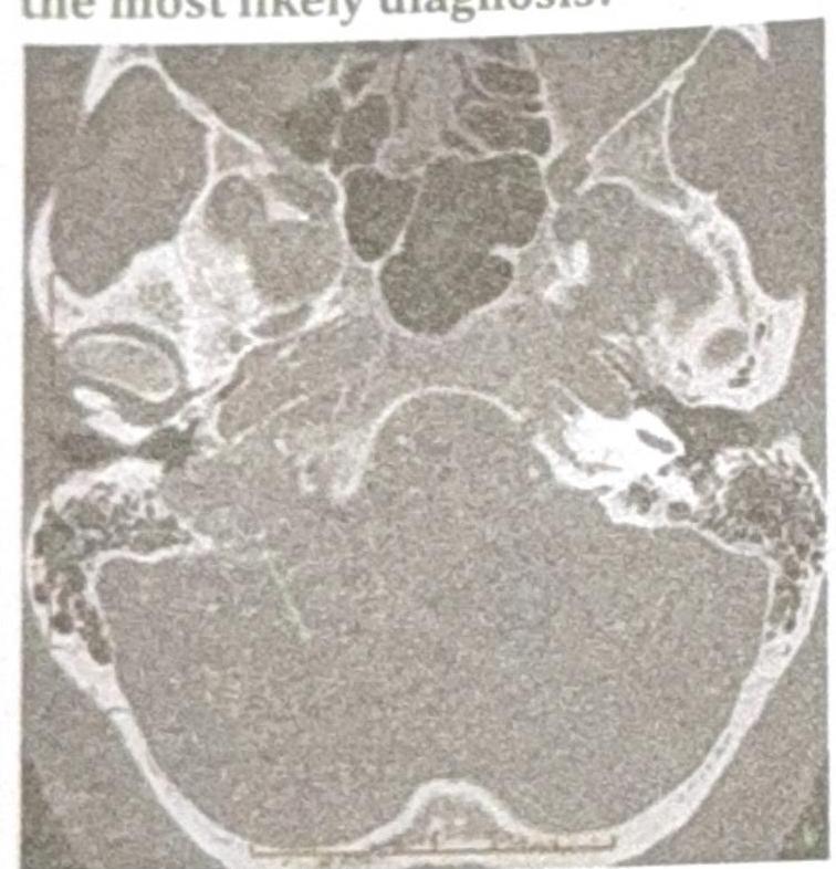
A patient presents with conductive hearing loss, pulsatile tinnitus, and a positive Phelps sign. Based on the CT scan image provided, what is the most likely diagnosis?
Q264
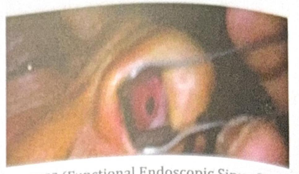
A patient with chronic nasal obstruction underwent a procedure 3 months ago and now presents with recurrent epistaxis, crusting, and the clinical image showing a septal perforation. What procedure was most likely carried out?