All (135)Anatomy (8)Anesthesiology (1)Biochemistry (9)Community Medicine (8)Dental (2)Dermatology (2)ENT (2)Forensic Medicine (3)Internal Medicine (20)Microbiology (7)Obstetrics and Gynecology (12)Ophthalmology (4)Orthopaedics (4)Pathology (6)Pediatrics (10)Pharmacology (13)Physiology (5)Psychiatry (2)Psychiatry (3)Radiology (1)Surgery (13)
Q121
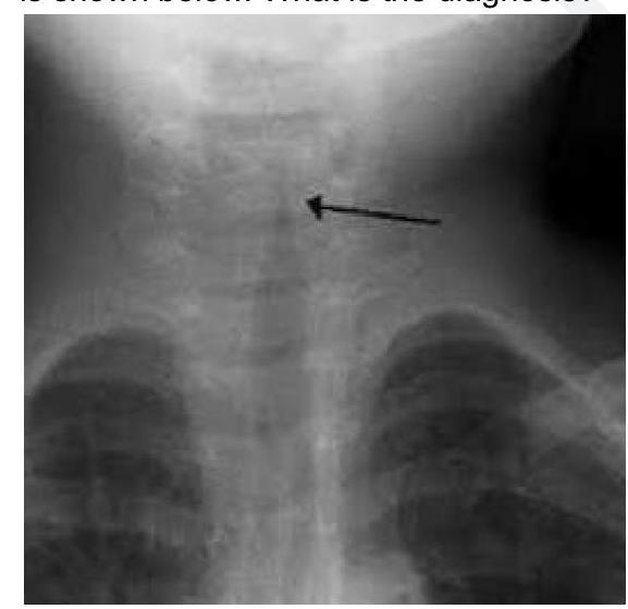
A 2 year child presented with low grade fever and stridor. What is the likely diagnosis?