NEET-PG 2021 — Radiology
6 Previous Year Questions with Answers & Explanations
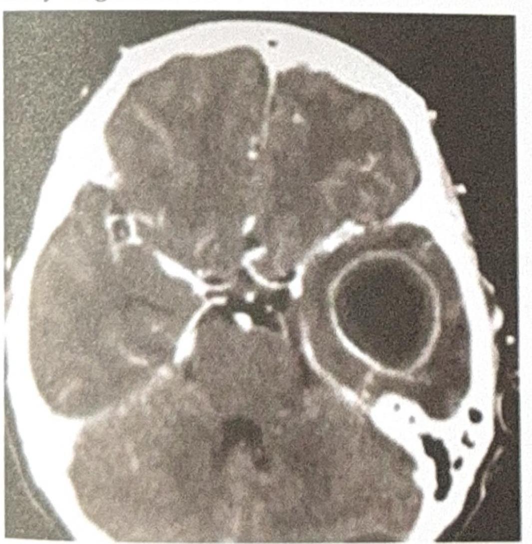
A patient with a history of chronic ear infection now presents with manifestations, including headache and vomiting. A CT brain image is shown. What is the most probable diagnosis?

Examine the abdominal X-ray shown. What is the most likely diagnosis based on the findings?
What is the modality of the test shown in the image?
A patient presents with abdominal distension. Based on the X-ray, which of the following bowel loops are dilated?
A 30-year-old female presents with sterile pyuria. The radiograph below is provided. What is the most likely diagnosis?

A patient presents with ear discharge. The CT image is shown below. Based on the clinical presentation and imaging, what is the most likely diagnosis?
NEET-PG 2021 - Radiology NEET-PG Practice Questions and MCQs
Question 1: A patient with a history of chronic ear infection now presents with manifestations, including headache and vomiting. A CT brain image is shown. What is the most probable diagnosis?
- A. Meningitis
- B. Extradural Abscess
- C. Cerebral Abscess
- D. Temporal lobe Abscess (Correct Answer)
Explanation: ***Temporal lobe Abscess*** - The CT scan shows a **ring-enhancing lesion** with significant surrounding edema, which is characteristic of a **brain abscess**. - Given the history of a **chronic ear infection**, the temporal lobe is a common site for bacterial spread from the mastoid air cells or middle ear. *Meningitis* - Meningitis involves inflammation of the **meninges** and typically presents with diffuse changes on imaging, such as sulcal effacement or leptomeningeal enhancement, rather than a focal, encapsulated lesion. - While it can cause headache and vomiting, the CT image does not show findings typical of meningitis. *Extradural Abscess* - An extradural (or epidural) abscess is located **between the dura mater and the skull bone**. - It would typically appear as a collection outside the brain parenchyma, potentially causing mass effect but distinct from an intraparenchymal lesion seen in the image. *Cerebral Abscess* - The image does show a **cerebral abscess**, but this option is less specific than "Temporal lobe abscess." - The question asks for the **most probable diagnosis**, and combining the imaging findings with the patient's history of ear infection points to a specific location within the cerebrum.
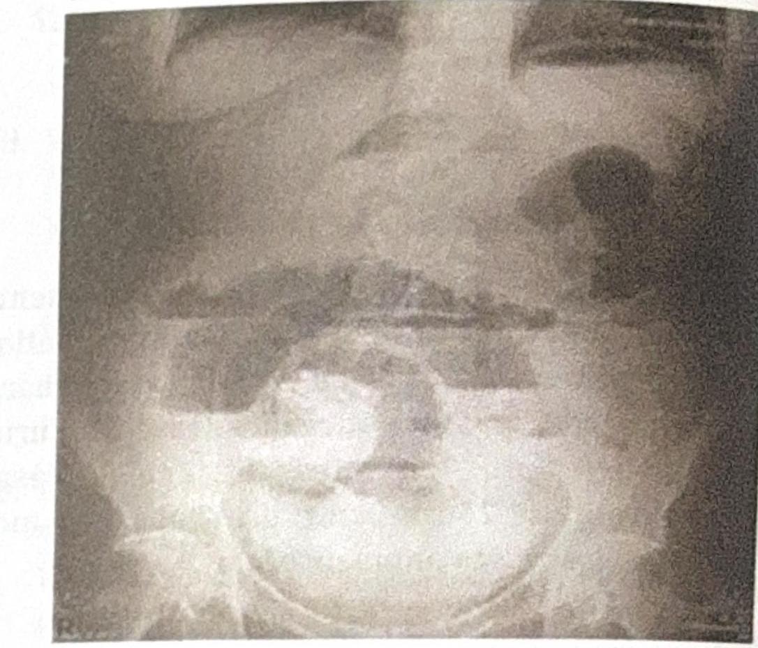
Question 2: Examine the abdominal X-ray shown. What is the most likely diagnosis based on the findings?
- A. Small bowel ileus
- B. Pneumoperitoneum
- C. Intestinal Obstruction (Correct Answer)
- D. Large bowel obstruction
Explanation: ***Intestinal Obstruction*** - The abdominal X-ray demonstrates **distended loops of bowel** with **multiple air-fluid levels**, which are classic radiographic signs of intestinal obstruction. - The presence of multiple, wide air-fluid levels visible in a **stepladder pattern** is a hallmark of bowel obstruction. - **Valvulae conniventes** (transverse folds crossing the entire width of bowel) suggest **small bowel** involvement when visible with distension. *Small bowel ileus* - While ileus can show distended bowel loops, it typically presents with **gas distributed throughout the small and large bowel** without a clear transition point. - Ileus shows **less pronounced air-fluid levels** and lacks the characteristic stepladder pattern seen in mechanical obstruction. - The clinical context and presence of multiple distinct air-fluid levels favor mechanical obstruction over ileus. *Large bowel obstruction* - Large bowel obstruction would show **dilated colon** with **haustrations** (incomplete folds that don't cross the entire lumen). - The obstruction would typically show dilation **proximal to the obstruction** with collapsed bowel distally. - The pattern in this image is more consistent with small bowel or generalized intestinal obstruction rather than isolated large bowel obstruction. *Pneumoperitoneum* - Pneumoperitoneum (free air in the peritoneal cavity) appears as **air under the diaphragm** on upright films or as **Rigler's sign** (both sides of bowel wall visible) on supine films. - This is a sign of **bowel perforation**, not obstruction with air-fluid levels within the bowel lumen. - The air-fluid levels seen here are **intraluminal**, not free intraperitoneal air.
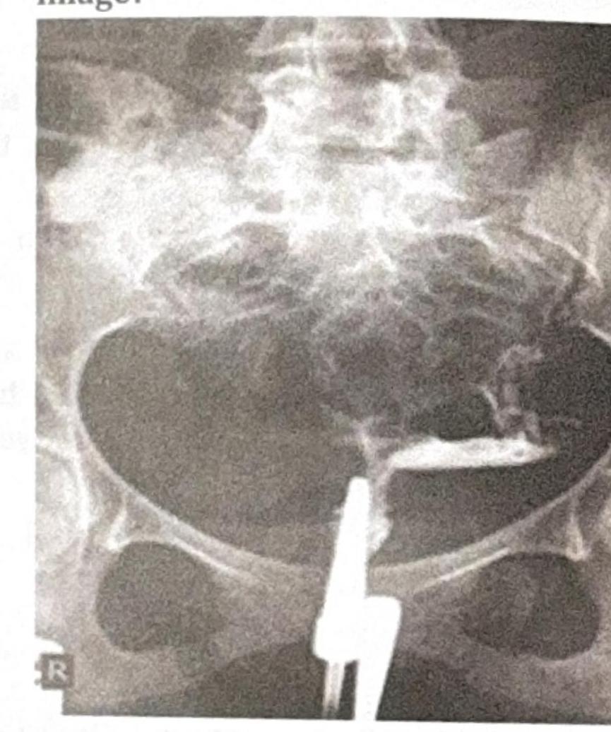
Question 3: What is the modality of the test shown in the image?
- A. Hysterosalpingography (Correct Answer)
- B. Hysteroscopy
- C. Laparoscopy
- D. Saline infusion sonography
Explanation: ***Hysterosalpingography*** - The image shows a **contrast-filled uterus and fallopian tubes**, characteristic of a **hysterosalpingogram (HSG)**. - An HSG uses **X-rays** and **radiopaque contrast media** to visualize the uterine cavity and assess fallopian tube patency. *Hysteroscopy* - **Hysteroscopy** involves direct visualization of the uterine cavity using a **fiber optic endoscope** inserted through the cervix. - It does not produce an X-ray image with contrast filling the fallopian tubes. *Laparoscopy* - **Laparoscopy** is a minimally invasive surgical procedure that involves inserting a **laparoscope** through an incision in the abdominal wall to view pelvic organs externally. - This image clearly depicts an internal view of the uterus and tubes through contrast, not an external, endoscopic view. *Saline infusion sonography* - **Saline infusion sonography (SIS)**, also known as sonohysterography, uses **ultrasound** imaging during the infusion of saline into the uterus. - While it assesses the uterine cavity, it is an ultrasound-based technique and does not involve X-ray contrast passing through the fallopian tubes, as seen in the image.
Question 4: A patient presents with abdominal distension. Based on the X-ray, which of the following bowel loops are dilated?
- A. Jejunum (Correct Answer)
- B. Duodenum
- C. Transverse colon
- D. Ileum
Explanation: ***Jejunum*** - The image shows dilated small bowel loops with prominent **valvulae conniventes** (also known as plicae circulares), which are characteristic of the jejunum. - These folds are typically closely spaced and extend across the entire lumen, giving a "coiled spring" or "stack of coins" appearance on plain radiographs when dilated. *Duodenum* - While the duodenum is part of the small bowel, it is the most proximal segment and typically not as diffusely involved in generalized small bowel dilation as the jejunum and ileum unless the obstruction is very high. - The valvulae conniventes in the duodenum are less prominent and more sparsely distributed compared to the jejunum. *Transverse colon* - The transverse colon is part of the large intestine and would show **haustra**, which are sacculations that do not extend across the entire lumen and are typically more widely spaced than valvulae conniventes. - The dilated loops in the image clearly show mucosal folds that span the entire width of the bowel. *Ileum* - The ileum also has valvulae conniventes, but they are less prominent and more sparsely distributed than in the jejunum. - In cases of small bowel obstruction or dilation, the jejunum characteristically shows more distinct and closely packed valvulae conniventes, making it the most identifiable segment in this image.
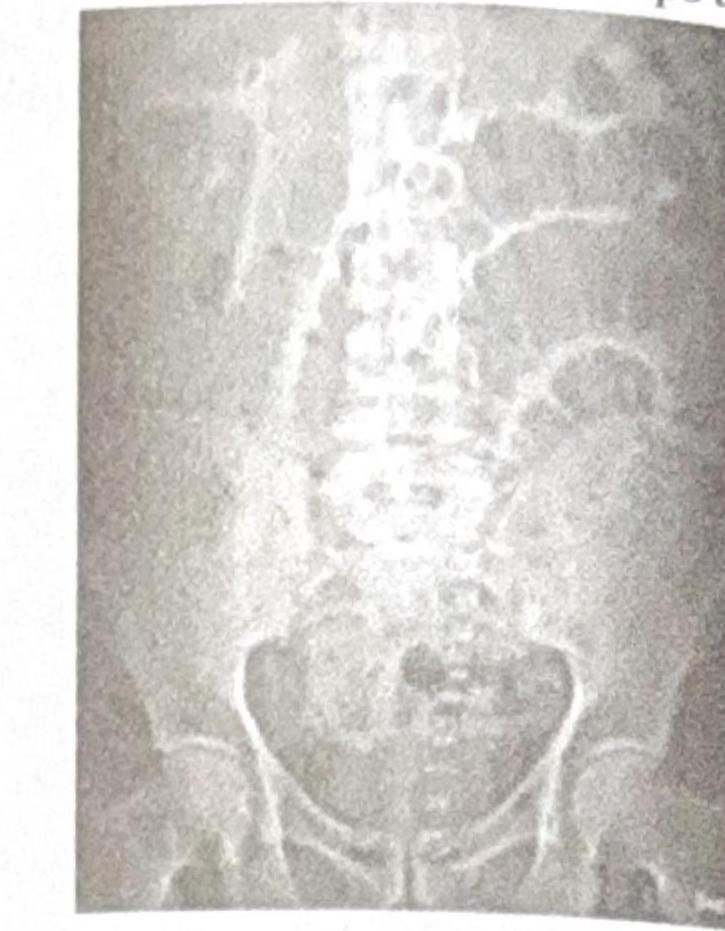
Question 5: A 30-year-old female presents with sterile pyuria. The radiograph below is provided. What is the most likely diagnosis?
- A. Psoas Calcification
- B. Putty Kidney (Correct Answer)
- C. Staghorn calculus
- D. Nephrocalcinosis
Explanation: ***Putty Kidney*** - The image visible in the question shows a **caseous calcification** of the renal parenchyma, which is characteristic of a "putty kidney," a late manifestation of **renal tuberculosis**. - **Sterile pyuria** is commonly associated with renal tuberculosis, where Mycobacterium tuberculosis infection leads to chronic inflammation and granuloma formation in the kidney, eventually resulting in caseous necrosis and calcification. *Psoas Calcification* - This typically refers to calcification within the **psoas muscle**, which would appear as a linear or amorphous calcification along the path of the muscle, an appearance not consistent with the image. - While psoas abscesses can calcify, they would not manifest as widespread renal parenchymal calcification and are not specifically linked to sterile pyuria in this manner. *Staghorn Calculus* - A **staghorn calculus** is a large, branched kidney stone that fills all or part of the renal pelvis and calyces, resembling the antlers of a stag. - While these stones are composed of mineral salts and would be radiopaque, their morphology is distinctly different from the diffuse, caseous calcification seen in the image. *Nephrocalcinosis* - **Nephrocalcinosis** is a condition characterized by diffuse microcalcifications throughout the renal parenchyma, usually affecting the tubules. - The calcifications in the provided image appear more nodular and clustered, consistent with late-stage tuberculous caseous necrosis, rather than the fine, diffuse pattern of nephrocalcinosis.
Question 6: A patient presents with ear discharge. The CT image is shown below. Based on the clinical presentation and imaging, what is the most likely diagnosis?
- A. Temporal lobe abscess (Correct Answer)
- B. Extradural abscess
- C. Cerebellar abscess
- D. Meningitis
Explanation: ***Temporal lobe abscess*** - The CT scan shows a **ring-enhancing lesion** in the **temporal lobe**, which is characteristic of a brain abscess. - **Ear discharge** (otorrhea), particularly from otitis media, is a common predisposing factor for temporal lobe abscesses due to the proximity of the middle ear and mastoid to the temporal lobe. - Otogenic brain abscesses account for a significant proportion of intracranial complications from ear infections, with the temporal lobe being the most common location. *Extradural abscess* - An **extradural abscess** would typically be located between the dura mater and the skull, often presenting as a **lenticular or biconvex collection** displacing the dura and brain, not within the brain parenchyma as seen here. - While ear infections can lead to extradural abscesses, the imaging clearly shows an intraparenchymal lesion. *Cerebellar abscess* - A **cerebellar abscess** would be located in the cerebellum (posterior fossa), which is a different anatomical location from the lesion seen in the image (which is in the supratentorial compartment, consistent with the temporal lobe). - Although ear infections can also lead to cerebellar abscesses, the lesion's position on the CT scan does not correspond to the cerebellum. *Meningitis* - **Meningitis** is an inflammation of the meninges and typically manifests on CT as **leptomeningeal enhancement**, particularly in the sulci and basal cisterns, rather than a discrete, encapsulated mass lesion like an abscess. - While ear discharge can be associated with meningitis, the imaging findings strongly point to an abscess, not diffuse meningeal inflammation.