All (182)Anatomy (10)Anatomy (1)Anesthesiology (3)Behavioral Science (1)Biochemistry (8)Biochemistry (1)Biostatistics (1)Community Medicine (8)Dermatology (4)ENT (4)General Medicine (1)Internal Medicine (14)Internal Medicine (10)Management (1)Microbiology (14)OB/GYN (11)Obstetrics and Gynecology (11)Ophthalmology (8)Orthopaedics (3)Pathology (6)Patient Safety (3)Pediatrics (1)Pediatrics (14)Pharmacology (18)Pharmacology (1)Physiology (6)Radiology (4)Surgery (15)
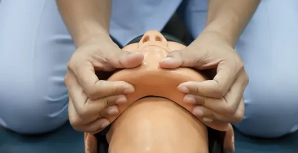
Q161
The procedure shown in the image is performed to maintain the airway. Which of the following techniques is being used?
Q162
A young male was administered regional anesthesia with 0.25% bupivacaine. The patient became unresponsive, and the pulse became unrecordable. What is the best management in this situation?
Q163
A patient is undergoing surgery where anesthesia is maintained with halothane. The patient develops hyperthermia and muscle rigidity. Which of the following agents is most likely responsible?