NEET-PG 2021 — Ophthalmology
6 Previous Year Questions with Answers & Explanations
What is the SI unit of illuminance (brightness of light on a surface)?
A patient with contact lens use for the past 2 years presents with the ocular findings shown in the image below. What is the most probable diagnosis?
A 15-year-old girl with myopic astigmatism does not want to wear glasses. What is the best alternative for her?
A one-month-old baby presents with excessive tearing (watering) and an increased corneal size. What is the most likely diagnosis?
An elderly woman presented with gradual painless diminution of vision. The fundus picture is shown below. What is the most likely diagnosis?
What is the most likely complication of the condition shown in the image below?
NEET-PG 2021 - Ophthalmology NEET-PG Practice Questions and MCQs
Question 1: What is the SI unit of illuminance (brightness of light on a surface)?
- A. Luminance
- B. Lux (Correct Answer)
- C. Candela
- D. Lumen
Explanation: ***Lux*** - **Lux** is the SI unit specifically designated for **illuminance**, which measures the **luminous flux** incident on a surface per unit area. - It quantifies the perceived **brightness** of light on a surface, taking into account the human eye's sensitivity to different wavelengths. *Luminance* - **Luminance** is a measure of the **intensity of light emitted or reflected from a surface** in a given direction, expressed in candelas per square meter (cd/m²). - It describes the brightness of a surface as perceived by the eye, but unlike illuminance, it is **independent of the incident light**. *Candela* - The **candela** is the SI base unit of **luminous intensity**, measuring the **power emitted by a light source in a particular direction**. - It doesn't describe the **brightness on a surface** but rather the output of the light source itself. *Lumen* - The **lumen** is the SI unit of **luminous flux**, representing the total amount of **visible light emitted by a source per unit time**. - While related to brightness, it describes the **total light output** of a source, not the illuminance on a specific surface.
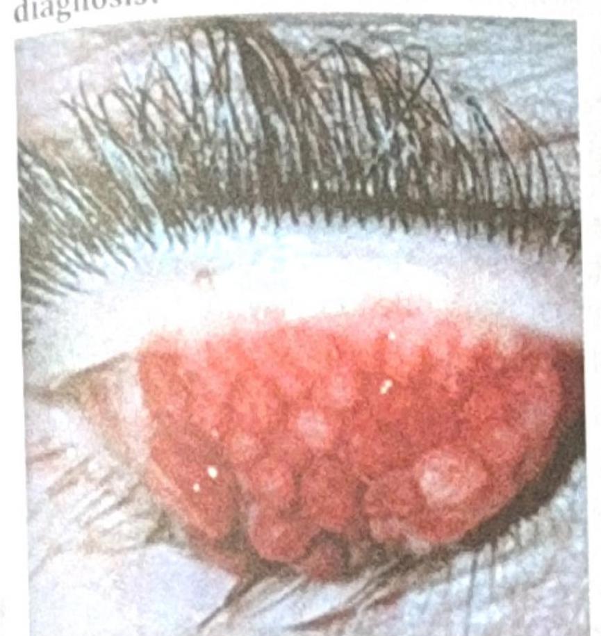
Question 2: A patient with contact lens use for the past 2 years presents with the ocular findings shown in the image below. What is the most probable diagnosis?
- A. Trachoma
- B. Giant Papillary conjunctivitis (Correct Answer)
- C. Ocular Surface Squamous Neoplasia (OSSN)
- D. Vernal Keratoconjunctivitis
Explanation: ***Giant Papillary conjunctivitis*** - The image shows **large, elevated papillae** on the **tarsal conjunctiva**, which are characteristic findings of giant papillary conjunctivitis. - This condition is common among **contact lens wearers**, caused by chronic mechanical irritation and an allergic response to lens material or deposits. *Trachoma* - Trachoma is a **chronic infectious disease** caused by *Chlamydia trachomatis*, leading to scarring of the conjunctiva. - It typically presents with **follicles** in the early stages, followed by **scarring** and **pannus formation**, not the large papillae seen here. *Ocular Surface Squamous Neoplasia (OSSN)* - OSSN refers to a spectrum of conditions from **dysplasia to squamous cell carcinoma** affecting the conjunctiva or cornea. - It usually presents as a **gelatinous, fleshy, or leukoplakic lesion**, often at the limbus, which is distinct from the diffuse papillae shown. *Vernal Keratoconjunctivitis* - Vernal keratoconjunctivitis (VKC) is a **severe form of allergic conjunctivitis** but primarily affects children and young adults with a history of atopy. - While it can cause large papillae (cobblestone papillae), it is not specifically associated with contact lens wear and usually has other systemic allergic manifestations.
Question 3: A 15-year-old girl with myopic astigmatism does not want to wear glasses. What is the best alternative for her?
- A. LASIK
- B. Spherical Specs
- C. Contact lenses (Toric) (Correct Answer)
- D. FEMTO Lasik
Explanation: ***Contact lenses (Toric)*** - **Toric contact lenses** are specifically designed to correct **astigmatism**, along with myopia or hyperopia, by having different refractive powers in different meridians. - They offer a non-surgical alternative to glasses, addressing the patient's desire not to wear spectacles, and are generally safe and effective for teenagers. *LASIK* - **LASIK (Laser-Assisted In Situ Keratomileusis)** is a surgical procedure to correct refractive errors, but it is not typically recommended for individuals under **18-21 years of age** due to continued eye growth and refractive changes. - The patient's age of 15 makes her an unsuitable candidate for LASIK at this time. *Spherical Specs* - **Spherical spectacles** are designed to correct myopia or hyperopia but cannot adequately correct **astigmatism**, which is a significant component of this patient's refractive error. - The patient also explicitly states she does not want to wear glasses, making this option undesirable. *FEMTO Lasik* - **FEMTO LASIK** is an advanced form of LASIK that uses a femtosecond laser to create the corneal flap, offering higher precision and safety. - However, similar to traditional LASIK, it is a **refractive surgical procedure** and typically not performed on patients younger than **18 years old** due to ongoing eye development.
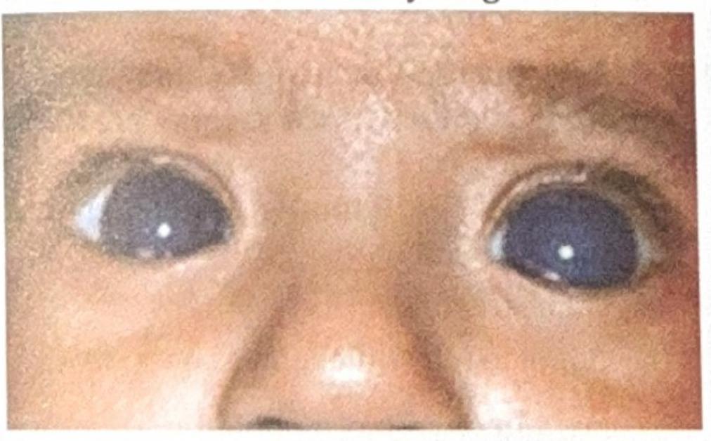
Question 4: A one-month-old baby presents with excessive tearing (watering) and an increased corneal size. What is the most likely diagnosis?
- A. Galactosemia
- B. Buphthalmos (Correct Answer)
- C. Cataract
- D. Hurler syndrome
Explanation: ***Buphthalmos*** - **Buphthalmos** refers to the enlargement of the eye in infants, typically caused by **congenital glaucoma**, which results in increased intraocular pressure. - The combination of **excessive tearing (epiphora)** and an **increased corneal size** (seen in the image as unusually large corneas for a one-month-old) are classic signs of buphthalmos due to elevated intraocular pressure stretching the infant's pliable sclera and cornea. *Galactosemia* - **Galactosemia** is a metabolic disorder that can cause cataracts and, in severe cases, liver damage and intellectual disability, but it does **not typically cause buphthalmos or enlarged corneas**. - While cataracts can lead to poor vision, they don't explain the excessive tearing or corneal enlargement. *Cataract* - A **cataract** is an opacity in the lens of the eye, which can cause blurry vision and a white pupil reflex (leukocoria), but **does not cause increased corneal size or excessive tearing** as primary symptoms. - While cataracts can occur in infants, they do not present with the specific combination of signs described. *Hurler syndrome* - **Hurler syndrome** is a lysosomal storage disorder (mucopolysaccharidosis type I) that can cause various ocular abnormalities, including **corneal clouding**, but typically **not corneal enlargement or buphthalmos**. - Other features include coarse facial features, skeletal abnormalities, and developmental delay, which are not mentioned in the presentation criteria.
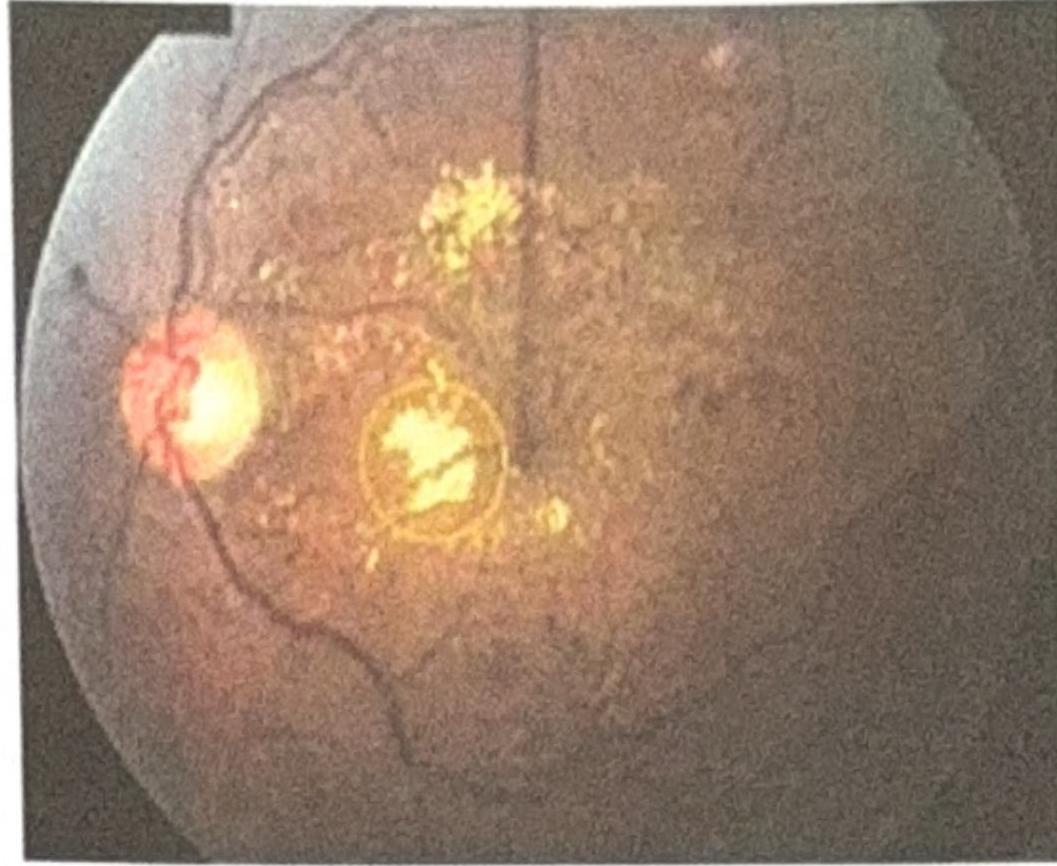
Question 5: An elderly woman presented with gradual painless diminution of vision. The fundus picture is shown below. What is the most likely diagnosis?
- A. Central Retinal Artery Occlusion (CRAO)
- B. Hypertensive Retinopathy
- C. Central Retinal Vein Occlusion (CRVO) (Correct Answer)
- D. Diabetic Retinopathy
Explanation: ***Central Retinal Vein Occlusion (CRVO)*** - The image displays characteristic findings of CRVO, including **widespread retinal hemorrhages**, **dilated and tortuous retinal veins**, and **cotton wool spots**. - The presence of **macular edema** (indicated by the bright, somewhat circular lesion near the center with surrounding exudates) also points to CRVO, which causes gradual, painless vision loss. *Central Retinal Artery Occlusion (CRAO)* - CRAO typically presents with sudden, profound, and painless vision loss, and the classic fundoscopic finding is a **cherry-red spot** in the macula with diffuse retinal whitening due to ischemia. - The image does not show these features; instead, it shows significant hemorrhages and dilated veins, which are inconsistent with CRAO. *Hypertensive Retinopathy* - Hypertensive retinopathy might show **arteriolar narrowing**, **AV nipping**, **cotton wool spots**, and sometimes hemorrhages, but the widespread, severe hemorrhages and marked venous dilation seen here are much more typical of CRVO. - While it can cause vision changes, the pattern of ocular findings is less severe and more chronic compared to the acute presentation of CRVO. *Diabetic Retinopathy* - Diabetic retinopathy can involve dot-blot hemorrhages, microaneurysms, hard exudates, and sometimes cotton wool spots, but the extensive, diffuse retinal hemorrhages in all four quadrants, along with the severely dilated and tortuous veins shown, are not the primary distinguishing features of typical diabetic retinopathy stages. - While **proliferative diabetic retinopathy (PDR)** can involve hemorrhages, the pattern in the image strongly suggests a vascular occlusion rather than the progressive microvascular damage of diabetes.
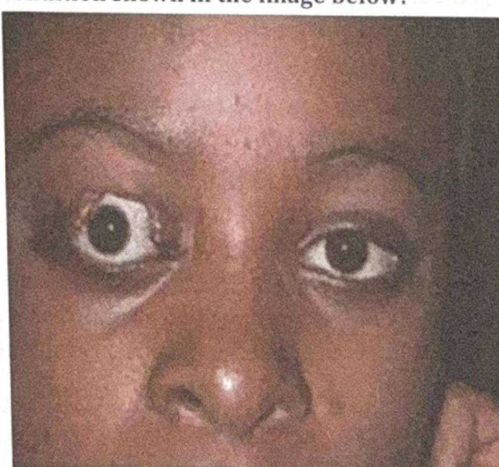
Question 6: What is the most likely complication of the condition shown in the image below?
- A. Exposure Keratitis (Correct Answer)
- B. Difficulty in eye movement
- C. Cataract
- D. Glaucoma
Explanation: ***Exposure Keratitis*** - The image shows **proptosis** (exophthalmos) of the right eye, where the eyeball protrudes forward. This condition often leads to incomplete eyelid closure (lagophthalmos). - **Exposure keratitis** occurs when the cornea is inadequately covered by the eyelids, leading to drying and damage due to constant exposure to air and environmental factors. *Difficulty in eye movement* - While **proptosis** can sometimes be associated with restricted eye movements (e.g., in severe Graves' ophthalmopathy due to muscle swelling), it is not the **most likely direct complication** of the exposure itself. - The image primarily depicts the physical displacement of the globe, which predisposes to corneal issues, not necessarily oculomotor dysfunction as the primary complication. *Cataract* - **Cataracts** are opacities of the lens and are typically associated with aging, trauma, or certain systemic conditions (e.g., diabetes, steroid use). - They are not a direct or common complication of **proptosis** or the resulting **exposure of the ocular surface**. *Glaucoma* - **Glaucoma** is a group of conditions characterized by damage to the optic nerve, often due to elevated intraocular pressure. - While severe **proptosis** leading to orbital congestion can theoretically increase intraocular pressure, it is not the most direct or prevalent complication compared to **exposure keratitis**, which is a direct consequence of inadequate globe protection.