NEET-PG 2021 — Microbiology
12 Previous Year Questions with Answers & Explanations
Name the parasite whose microfilariae have a sheath and no nuclei at the tail end.
A patient presents with a history of pastry intake causing food poisoning. What is the most likely causative agent?
A patient presented with 70% burns, and a sample was collected from the burn site. The image shows Gram-negative rods, and the suspected organism is an obligate aerobe. What is the most likely causative microbe?
A truck driver presented with a painless, demarcated ulcer on the penis and inguinal lymphadenopathy. What is the best method to visualize the motility of the most likely causative agent?
A patient was suspected of having brucellosis. A serum sample was sent for a standard agglutination test, which was initially negative but became positive after dilution of the sample. What is the most likely reason for the initial negative test?
A patient on steroids presented with nocturnal cough and chronic urticaria. Bronchoalveolar lavage (BAL) staining was done, and the organism shown in the image was identified. What is the most likely organism?
A child presented with bloody stools and abdominal pain. Which enrichment medium should be used for processing the fecal sample?
What is the vector for Leishmania, a parasite characterized by a prominent kinetoplast in its morphological forms?
A child presented with bluish-white spots in the mouth followed by a rash. What is the genome of the most likely causative agent?
A group of people ate patty late at night and experienced bouts of vomiting early in the morning. What is the most likely cause?
NEET-PG 2021 - Microbiology NEET-PG Practice Questions and MCQs
Question 1: Name the parasite whose microfilariae have a sheath and no nuclei at the tail end.
- A. Brugia malayi
- B. Loa loa
- C. Onchocerca volvulus
- D. Wuchereria bancrofti (Correct Answer)
Explanation: **Wuchereria bancrofti** - **Wuchereria bancrofti** microfilariae are characterized by the presence of a **sheath** and a **clear tail end** that is devoid of nuclei. - This morphology is a key feature used in the microscopic differentiation of W. bancrofti from other filarial species. *Brugia malayi* - **Brugia malayi** microfilariae also have a **sheath**, but their tail end typically contains **two distinct nuclei** that are spaced apart. - They are generally shorter and have more kinky curves than W. bancrofti. *Loa loa* - **Loa loa** microfilariae possess a **sheath** but are distinguished by their **nuclei extending to the tip** of the tail, often in a more continuous pattern. - They are also known for their diurnal periodicity, which aids in diagnosis. *Onchocerca volvulus* - **Onchocerca volvulus** microfilariae are **unsheathed** and have nuclei that do **not extend to the tail tip**, leaving a distinct clear space. - They are typically found in the skin and subcutaneous tissue, rather than the blood.
Question 2: A patient presents with a history of pastry intake causing food poisoning. What is the most likely causative agent?
- A. Verotoxin-producing E. coli
- B. Bacillus cereus
- C. Staphylococcus aureus (Correct Answer)
- D. Enteroinvasive E. coli (EIEC)
Explanation: ***Staphylococcus aureus*** - *S. aureus* is a common cause of food poisoning linked to **creamy foods** like pastries, salads, and custards, as it produces **heat-stable enterotoxins** when allowed to proliferate. - The symptoms, typically rapid onset **nausea, vomiting**, and abdominal cramps, occur because of the **preformed toxins** in the food, not necessarily active infection. *Verotoxin-producing E. coli* - This strain, often **E. coli O157:H7**, is primarily associated with **undercooked beef** or contaminated produce, and typically causes **bloody diarrhea** and can lead to hemolytic uremic syndrome (HUS). - Its mechanism involves **verotoxins** directly damaging intestinal cells and blood vessels, which is different from the rapid, emetic-focused symptoms of *S. aureus* food poisoning. *Bacillus cereus* - *B. cereus* causes two main types of food poisoning: **emetic (vomiting)**, typically from **reheated rice**, and **diarrheal**, from meat products or vegetables. - While the emetic form can cause vomiting, it is most strongly associated with **rice dishes** and usually has a shorter incubation period than the diarrheal form, making *Staphylococcus aureus* a more classic cause for pastry-related outbreaks. *Enteroinvasive E. coli (EIEC)* - EIEC causes a disease similar to **shigellosis**, involving direct invasion of intestinal epithelial cells, leading to **bloody diarrhea** and fever. - It is typically spread through contaminated food and water and not specifically linked to pastry intake or characterized by the rapid onset emetic symptoms seen with preformed toxins.
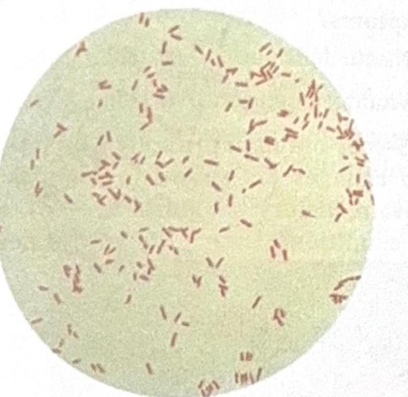
Question 3: A patient presented with 70% burns, and a sample was collected from the burn site. The image shows Gram-negative rods, and the suspected organism is an obligate aerobe. What is the most likely causative microbe?
- A. Neisseria meningitidis (Meningococcus)
- B. Streptococcus pneumoniae (Pneumococcus)
- C. Pseudomonas aeruginosa (Correct Answer)
- D. Streptococcus pyogenes
Explanation: ***Pseudomonas aeruginosa*** - The image shows **Gram-negative rods**, and the patient has extensive **burns**, making *Pseudomonas aeruginosa* a highly likely causative agent due to its common association with burn wound infections. - *Pseudomonas aeruginosa* is an **obligate aerobe** and thrives in moist environments, making it a frequent colonizer of burn wounds, which are large, often moist surfaces. *Neisseria meningitidis (Meningococcus)* - *Neisseria meningitidis* is a **Gram-negative coccus**, typically appearing as diplococci, not rods, on Gram stain. - While it can cause severe infections, it is primarily associated with **meningitis** and **sepsis**, not typically burn wound infections. *Streptococcus pneumoniae (Pneumococcus)* - *Streptococcus pneumoniae* is a **Gram-positive coccus**, appearing as lancet-shaped diplococci or short chains, which contradicts the Gram-negative rod morphology seen in the image. - It is a common cause of **pneumonia** and **otitis media**, not primarily associated with burn wound infections. *Streptococcus pyogenes* - *Streptococcus pyogenes* is a **Gram-positive coccus** that grows in chains, which is inconsistent with the Gram-negative rod morphology. - Although it can cause skin infections like cellulitis and impetigo, it is not a typical cause of **burn wound infections** in the way *Pseudomonas aeruginosa* is.
Question 4: A truck driver presented with a painless, demarcated ulcer on the penis and inguinal lymphadenopathy. What is the best method to visualize the motility of the most likely causative agent?
- A. Fluorescent microscopy
- B. Light microscopy
- C. Dark field microscopy (Correct Answer)
- D. Electron microscopy
Explanation: ***Dark field microscopy*** - The symptoms (painless, demarcated penile ulcer and inguinal lymphadenopathy) are highly suggestive of **primary syphilis**, caused by *Treponema pallidum*. - **Dark field microscopy** is the gold standard for visualizing the characteristic **corkscrew motility** of *T. pallidum* directly from lesion exudate. *Fluorescent microscopy* - This technique uses **fluorochromes** to stain structures and is often used in **immunofluorescence** assays to detect antibodies or antigens. - While useful for some microbial identification, it is not the primary method for visualizing the motility of *Treponema pallidum*. *Light microscopy* - Standard light microscopy has **insufficient resolution** to clearly visualize the thin, coiled spirochetes of *Treponema pallidum* or their motility. - The organisms are generally **too small and refractile** to be easily seen without specialized illumination. *Electron microscopy* - Provides extremely **high resolution** and is used for studying viral structures or detailed cellular ultrastructure. - It is **not practical** for routine clinical diagnosis, especially for live, motile bacteria, and is not used to observe motility.
Question 5: A patient was suspected of having brucellosis. A serum sample was sent for a standard agglutination test, which was initially negative but became positive after dilution of the sample. What is the most likely reason for the initial negative test?
- A. Antigen antibody complexes
- B. Postzone phenomenon
- C. Complement inactivation
- D. Prozone phenomenon (Correct Answer)
Explanation: ***Correct: Prozone phenomenon*** - The **prozone phenomenon** occurs when there is a very high concentration of antibodies in the patient's serum, leading to the formation of small antigen-antibody complexes that do not agglutinate or precipitate. - Diluting the sample reduces the antibody concentration, allowing for optimal antigen-antibody lattice formation and visible agglutination. - This is the classic explanation for a **negative test becoming positive after dilution** in brucellosis serology. *Incorrect: Antigen antibody complexes* - While agglutination tests rely on the formation of **antigen-antibody complexes**, the initial negative result despite a positive finding after dilution indicates a specific issue with complex *visibility* or *stability* rather than the general presence of complexes. - This option is too general and doesn't explain why dilution would change the result from negative to positive. *Incorrect: Postzone phenomenon* - The **postzone phenomenon** occurs when there is an *excess of antigen* relative to antibody, leading to no visible agglutination. - In such a case, diluting the sample (which would reduce antigen concentration or keep antibody concentration too low) would typically *not* lead to a positive result; in fact, further dilution of antibodies would worsen the outcome. - Postzone is the opposite mechanism and would not be corrected by dilution. *Incorrect: Complement inactivation* - **Complement inactivation** is not directly relevant to the mechanism of agglutination tests, which primarily depend on direct antibody-antigen binding for visible clumping. - These tests do not typically require complement activity for their primary reaction, nor are they inhibited by complement inactivation.
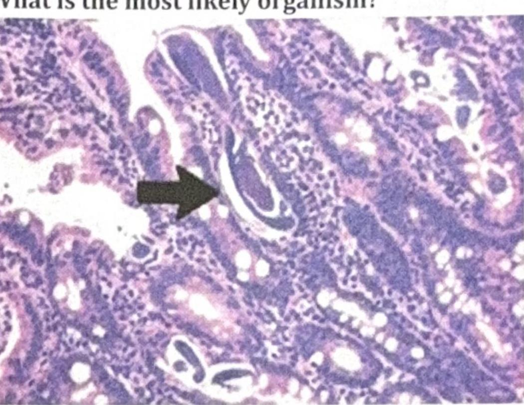
Question 6: A patient on steroids presented with nocturnal cough and chronic urticaria. Bronchoalveolar lavage (BAL) staining was done, and the organism shown in the image was identified. What is the most likely organism?
- A. Enterobius vermicularis
- B. Strongyloides stercoralis (Correct Answer)
- C. Capillaria philippinensis
- D. Ancylostoma duodenale
Explanation: ***Strongyloides stercoralis*** - The image shows a nematode larva in a **bronchoalveolar lavage (BAL) sample**, consistent with **Strongyloides stercoralis**, which is known for its ability to **autoinfect** and cause chronic infections. - The patient's **steroid use** can lead to **Strongyloides hyperinfection syndrome**, explaining the pulmonary symptoms (**nocturnal cough**) due to larval migration into the lungs and the chronic **urticaria** often associated with strongyloidiasis. *Enterobius vermicularis* - This organism primarily causes **pinworm infection**, characterized by **perianal itching** and is typically identified by the **scotch tape test** for eggs, not usually found in BAL or associated with chronic urticaria. - It does not commonly cause significant pulmonary symptoms or hyperinfection syndrome, especially not triggered by steroid use. *Capillaria philippinensis* - This nematode causes **intestinal capillariasis**, characterized by **chronic diarrhea**, **abdominal pain**, and **malabsorption**. - It is not typically associated with pulmonary symptoms like nocturnal cough or chronic urticaria, and is transmitted through fish consumption. *Ancylostoma duodenale* - This is a **hookworm** that causes **iron deficiency anemia**. While it does have a lung migration phase that can cause transient cough, it is not typically associated with chronic urticaria or severe pulmonary disease due to steroid-induced hyperinfection. - The image shows larvae, but the clinical context strongly points to *Strongyloides* due to the steroid use and specific symptoms.
Question 7: A child presented with bloody stools and abdominal pain. Which enrichment medium should be used for processing the fecal sample?
- A. Blood agar
- B. Selenite F broth (Correct Answer)
- C. Alkaline peptone water
- D. Muller Hinton Broth
Explanation: ***Selenite F broth*** - This **enrichment medium** is specifically designed to isolate **Salmonella** and some species of **Shigella**, which are common causes of bloody stools and abdominal pain in children. - It inhibits the growth of commensal gut flora, allowing pathogenic bacteria to proliferate and be subsequently identified on selective media. *Blood agar* - Blood agar is a **general-purpose enrichment medium** that supports the growth of a wide range of bacteria but does not selectively enrich for specific pathogens. - It would be ineffective in outcompeting the normal fecal flora to isolate rarer enteric pathogens causing the symptoms. *Alkaline peptone water* - This medium is primarily used for the enrichment of **Vibrio cholerae** species, which typically cause watery diarrhea, not bloody stools. - While it helps in the isolation of *Vibrio* species, it is not suitable for the suspected pathogens in this clinical scenario. *Muller Hinton Broth* - Muller-Hinton media are primarily used for **antimicrobial susceptibility testing** (antibiotic sensitivity testing) and are not designed for the primary isolation or enrichment of specific pathogens from clinical samples. - It would not provide a selective advantage for the recovery of organisms causing bloody diarrhea from a fecal sample.
Question 8: What is the vector for Leishmania, a parasite characterized by a prominent kinetoplast in its morphological forms?
- A. Female Anopheles Mosquito
- B. Triatomine bug
- C. Sand fly (Correct Answer)
- D. TseTse fly
Explanation: ***Sand fly*** - **Sand flies** (genus *Phlebotomus* in the Old World and *Lutzomyia* in the New World) are the biological vectors responsible for transmitting *Leishmania* parasites to humans and other mammals. - The parasite exists in two main forms; the **promastigote** (flagellated) in the sand fly gut and the **amastigote** (non-flagellated) within host macrophages. *Female Anopheles Mosquito* - The **female Anopheles mosquito** is the primary vector for **malaria**, caused by *Plasmodium* parasites, and not *Leishmania*. - *Plasmodium* also undergoes sporogonic development in the mosquito, but it does not have a prominent kinetoplast in its mature forms. *Triatomine bug* - The **triatomine bug**, also known as the "kissing bug", is the vector for **Chagas disease**, caused by *Trypanosoma cruzi*. - While *Trypanosoma cruzi* is also a hemoflagellate with a kinetoplast, it is associated with different disease manifestations and geographical distribution than *Leishmania*. *TseTse fly* - The **tsetse fly** (genus *Glossina*) is the vector responsible for transmitting **African Trypanosomiasis**, or sleeping sickness, caused by *Trypanosoma brucei*. - Like *Leishmania*, *Trypanosoma brucei* also possesses a kinetoplast, but it causes a clinically distinct disease and is transmitted by a different insect vector.
Question 9: A child presented with bluish-white spots in the mouth followed by a rash. What is the genome of the most likely causative agent?
- A. Enveloped virus with single-stranded RNA (Correct Answer)
- B. Double stranded Naked RNA
- C. Naked virus with single-stranded RNA
- D. Double stranded Enveloped RNA
Explanation: ***Enveloped virus with single-stranded RNA*** - Bluish-white spots in the mouth (**Koplik spots**) followed by a rash are pathognomonic for **measles**, which is caused by the **measles virus**. - The measles virus is a **paramyxovirus**, characterized as an **enveloped, negative-sense, single-stranded RNA virus**. *Double stranded Naked RNA* - No major human pathogen belongs to this specific genomic and structural classification. - Most **dsRNA viruses** like **rotavirus** are **naked** but cause gastroenteritis, not measles. *Naked virus with single-stranded RNA* - Viruses like **rhinovirus** (common cold) or **poliovirus** fit this description but do not cause Koplik spots or measles. - **Naked viruses** lack a lipid envelope, making them generally more resistant to environmental factors. *Double stranded Enveloped RNA* - There are no known medically significant human viruses that are both **enveloped** and contain **double-stranded RNA**. - Viral genomes are typically either DNA or RNA, and RNA viruses are usually single-stranded (positive or negative sense) or double-stranded, with or without an envelope.
Question 10: A group of people ate patty late at night and experienced bouts of vomiting early in the morning. What is the most likely cause?
- A. Escherichia coli
- B. Staphylococcus aureus (Correct Answer)
- C. Lactobacillus
- D. Bacillus cereus
Explanation: ***Staphylococcus aureus*** - This scenario describes classic **Staphylococcal food poisoning** with rapid onset of vomiting (4-8 hours after ingestion). - *S. aureus* produces preformed **heat-stable enterotoxins** in contaminated foods left at room temperature, particularly **meat products, pastries, and cream-filled items**. - Clinical hallmark: **Prominent vomiting** with minimal diarrhea and rapid resolution (24 hours). - The timing (late night eating → early morning vomiting) and food item (patty) make this the **definitive diagnosis**. *Escherichia coli* - Most pathogenic *E. coli* strains (ETEC, EHEC, EPEC) have **longer incubation periods** (1-3 days). - Typically presents with **diarrhea as the predominant symptom**, not isolated vomiting. - The rapid onset of symptoms excludes direct bacterial infection and indicates preformed toxin ingestion. *Lactobacillus* - These are **probiotic bacteria** and part of normal human flora (gut, vagina, mouth). - Not pathogenic and not associated with food poisoning. - Used therapeutically to prevent antibiotic-associated diarrhea. *Bacillus cereus* - While *B. cereus* has an emetic form with rapid onset (1-5 hours), it is **classically associated with reheated fried rice** and starchy foods, not meat products. - The emetic toxin (cereulide) is produced in improperly stored rice, not patties. - The food item (patty) clearly points to *S. aureus* rather than *B. cereus*.