All SubjectsAnatomy (27)Anesthesiology (4)Biochemistry (28)Biochemistry (1)Community Medicine (12)Dental (1)Dermatology (8)ENT (4)Forensic Medicine (3)General Medicine (3)Internal Medicine (41)Microbiology (25)Obstetrics and Gynecology (24)Ophthalmology (3)Orthopaedics (6)Pathology (35)Pathology (4)Pediatrics (22)Pharmacology (23)Physiology (13)Psychiatry (8)Psychiatry (3)Radiology (26)Surgery (8)Surgery (34)
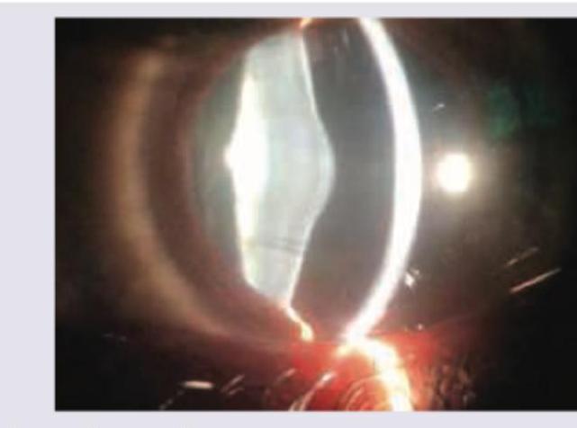
Q11
A child during evaluation of recurrent hematuria has the following eye finding. He has sensorineural deafness and history of similar illness in family members. What is the diagnosis?
Q12
Which is true about an infant with failure to thrive and the following findings?
Q13
This baby has obesity along with organomegaly. Comment on the diagnosis from the hint given in the image?
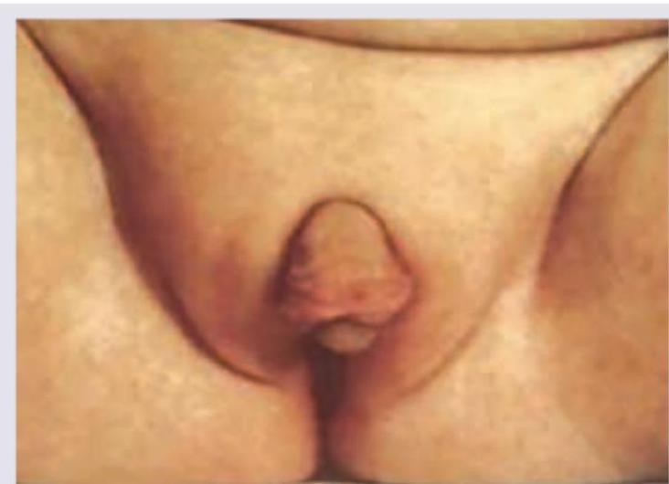
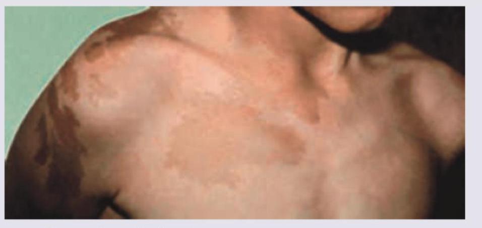
Q14
A 5-year-old girl is referred for vaginal bleeding. Physical examination shows breast development, multiple cystic changes in bone radiologically and following skin finding. What is the most likely pubertal disorder?
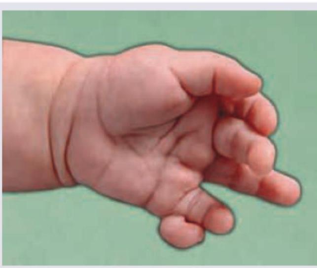
Q15
During evaluation of a child with obesity, following finding is observed. What is the association?
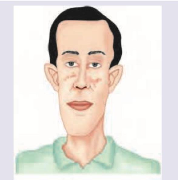
Q16
During evaluation of intellectual disability, characteristic facies of the child as below is noted. There is a family history of intellectual disability also. What is the probable diagnosis?
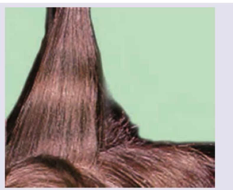
Q17
Which of the following is associated with this condition?
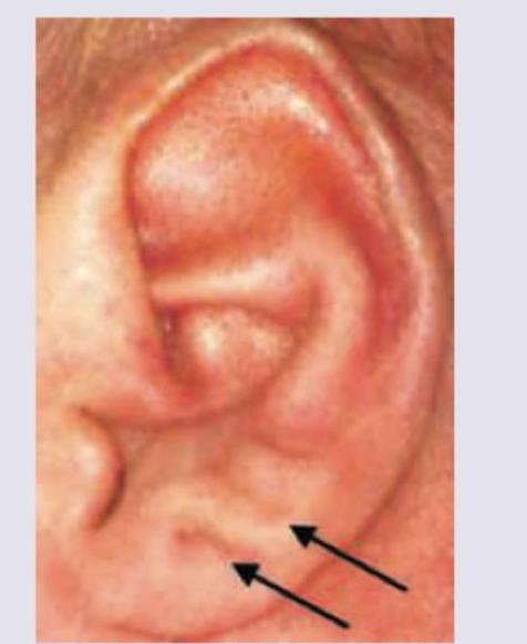
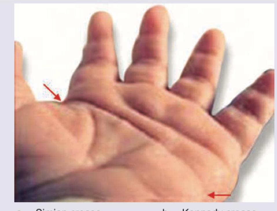
Q18
During evaluation of a child with Down syndrome, the following finding is noted. Identify?
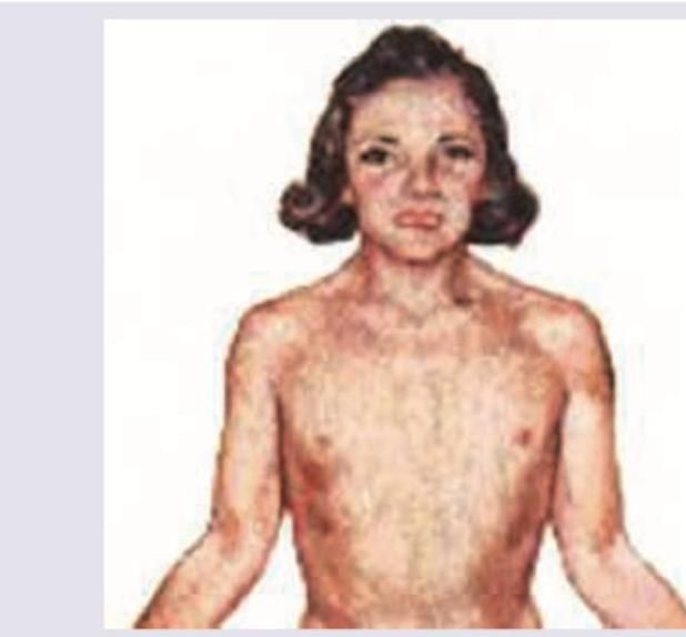
Q19
What is the expected karyotype in this child with the following findings and having pulmonic stenosis on Echocardiography?
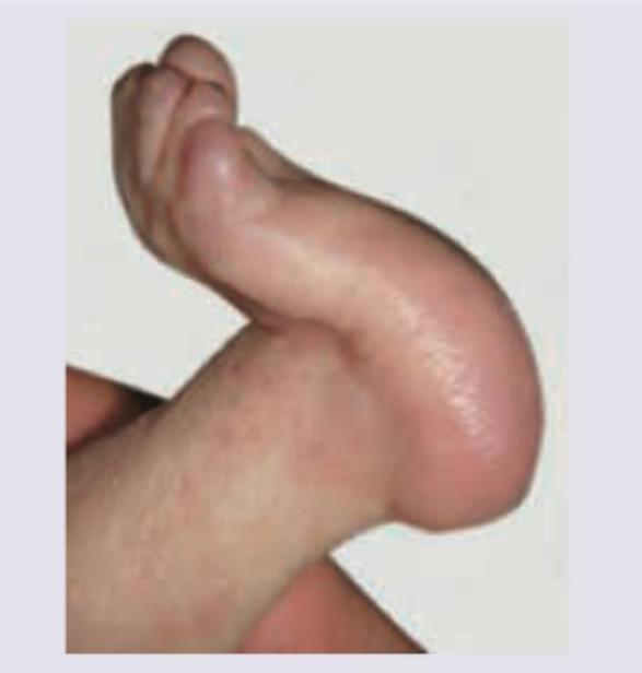
Q20
Which condition is characterized by the sign shown in the image?