All (366)Anatomy (27)Anesthesiology (4)Biochemistry (28)Biochemistry (1)Community Medicine (12)Dental (1)Dermatology (8)ENT (4)Forensic Medicine (3)General Medicine (3)Internal Medicine (41)Microbiology (25)Obstetrics and Gynecology (24)Ophthalmology (3)Orthopaedics (6)Pathology (35)Pathology (4)Pediatrics (22)Pharmacology (23)Physiology (13)Psychiatry (8)Psychiatry (3)Radiology (26)Surgery (8)Surgery (34)
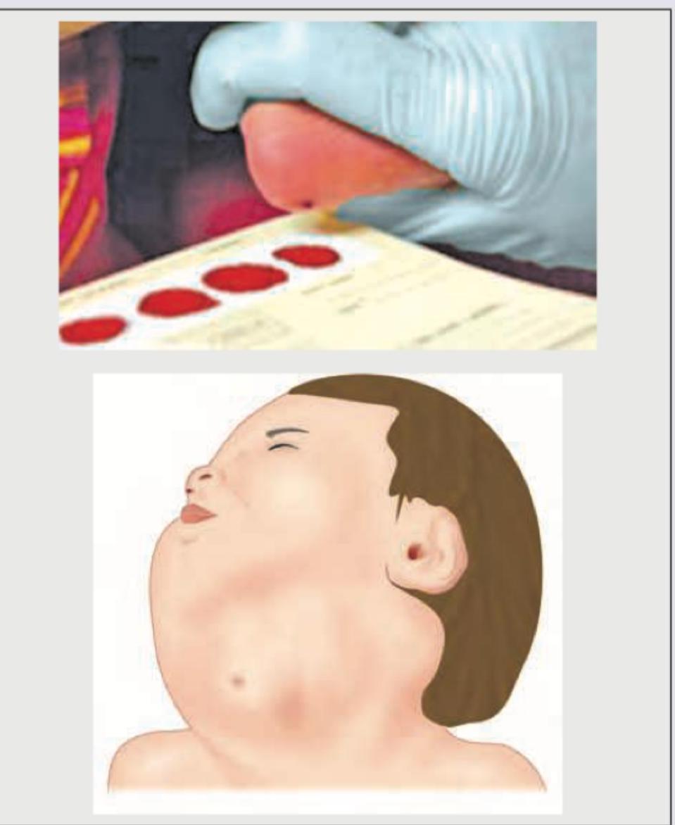
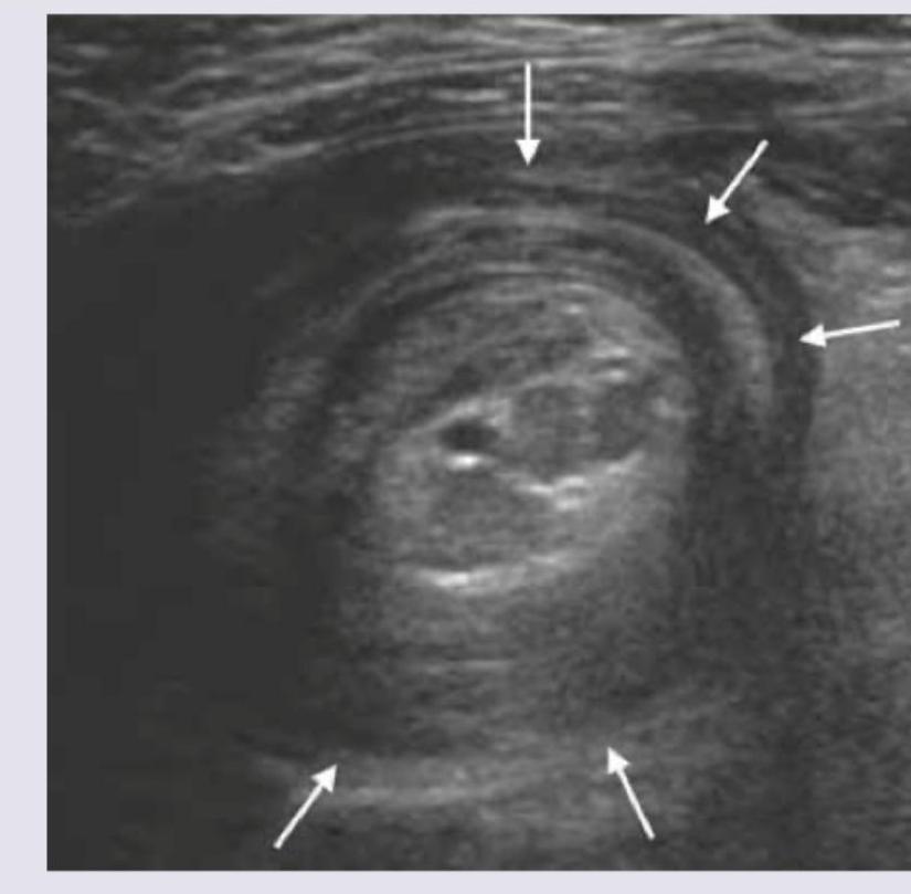
Q321
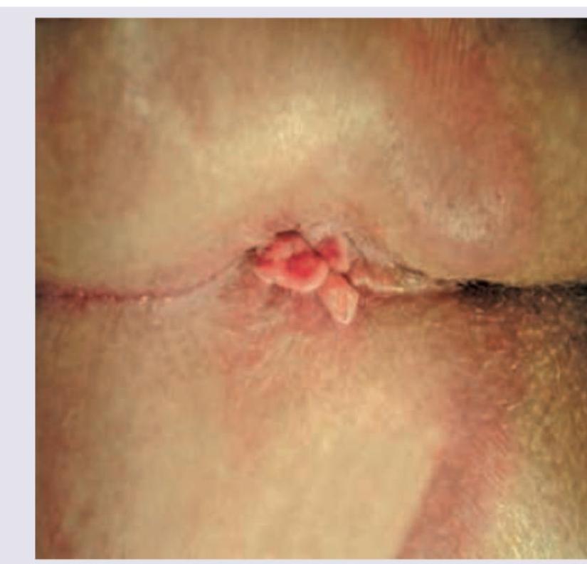
A 15-year-old boy presents with abdominal cramps and diarrhea for 8 weeks along with delayed puberty. Perianal examination reveals the following. What is the diagnosis? (NEET Pattern 2018)
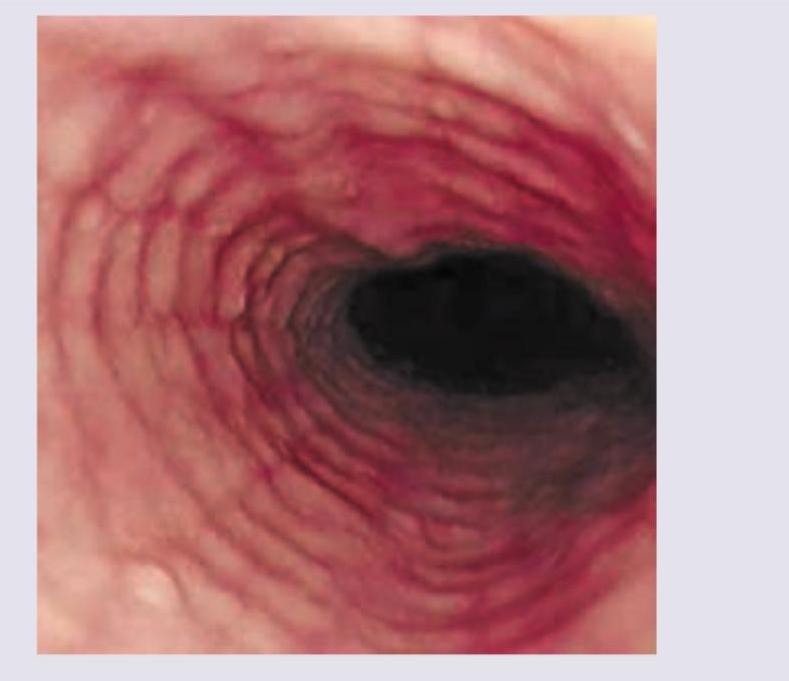
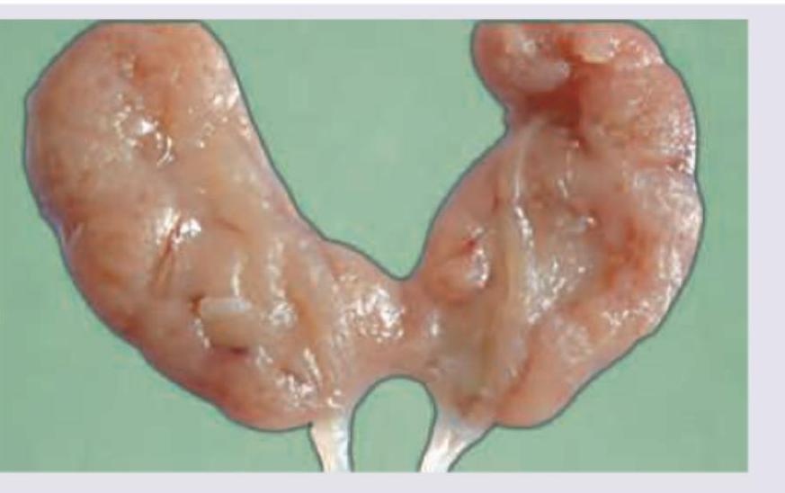
Q322
The given endoscopy of the patient shows? (NEET Pattern 2018)