All SubjectsAnatomy (27)Anesthesiology (4)Biochemistry (28)Biochemistry (1)Community Medicine (12)Dental (1)Dermatology (8)ENT (4)Forensic Medicine (3)General Medicine (3)Internal Medicine (41)Microbiology (25)Obstetrics and Gynecology (24)Ophthalmology (3)Orthopaedics (6)Pathology (35)Pathology (4)Pediatrics (22)Pharmacology (23)Physiology (13)Psychiatry (8)Psychiatry (3)Radiology (26)Surgery (8)Surgery (34)
Q11
Sabin Feldman dye test is used for diagnosis of which of the following condition?
Q12
Which of the following is the carrying agent for Lyme disease?
Q13
Urea breath test is used for diagnosis of:
Q14
Whole blood is used as a sample for which of the following tests?
Q15
Most common catheter-related bloodstream infection is due to:
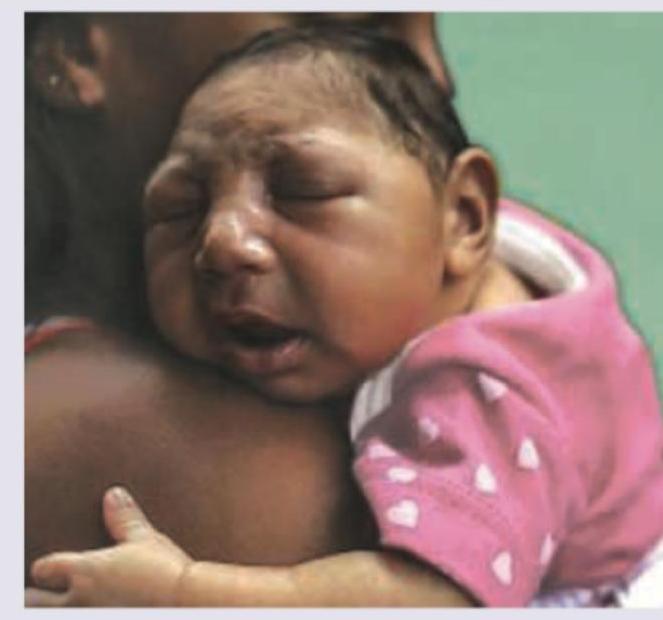
Q16
Which of the following is responsible for the abnormality shown below?
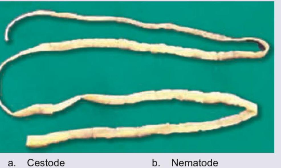
Q17
The following organism is called:
Q18
Which of the following life cycle is shown below?
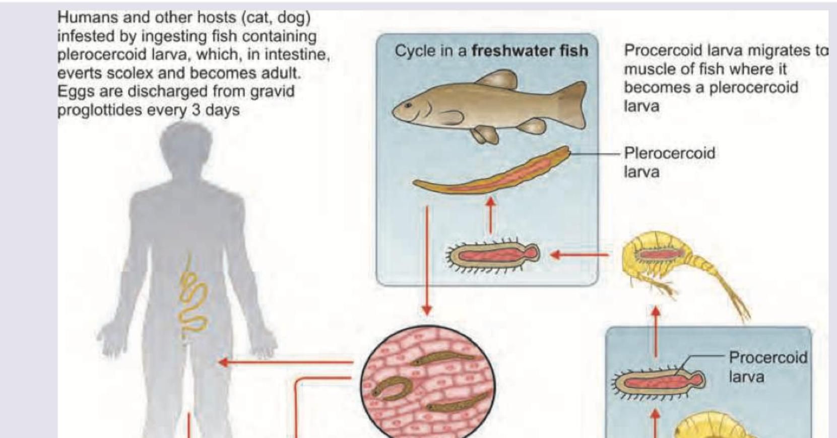
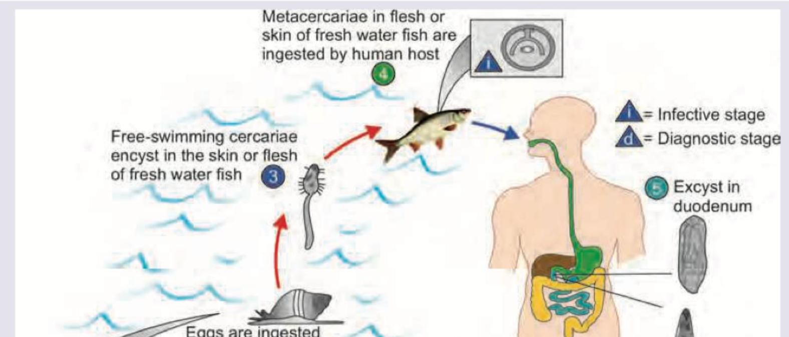
Q19
Which of the following life cycle is shown below?
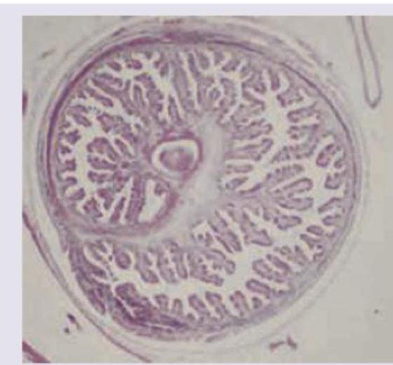
Q20
Which of the following parasite is shown below?