All SubjectsAnatomy (27)Anesthesiology (4)Biochemistry (28)Biochemistry (1)Community Medicine (12)Dental (1)Dermatology (8)ENT (4)Forensic Medicine (3)General Medicine (3)Internal Medicine (41)Microbiology (25)Obstetrics and Gynecology (24)Ophthalmology (3)Orthopaedics (6)Pathology (35)Pathology (4)Pediatrics (22)Pharmacology (23)Physiology (13)Psychiatry (8)Psychiatry (3)Radiology (26)Surgery (8)Surgery (34)
Q21
During acute tonsillitis, referred pain from the tonsil to the middle ear occurs via which nerve?
Q22
Testicular artery is a branch of -
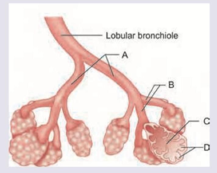
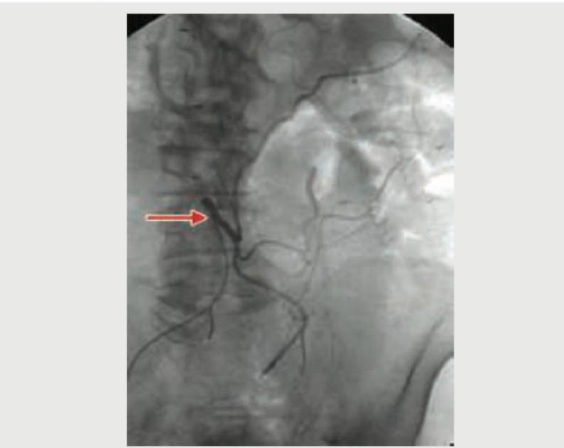
Q23
The Gut blood vessel marked (Red arrow) in the angiogram is: (Recent NEET Pattern 2018-19)
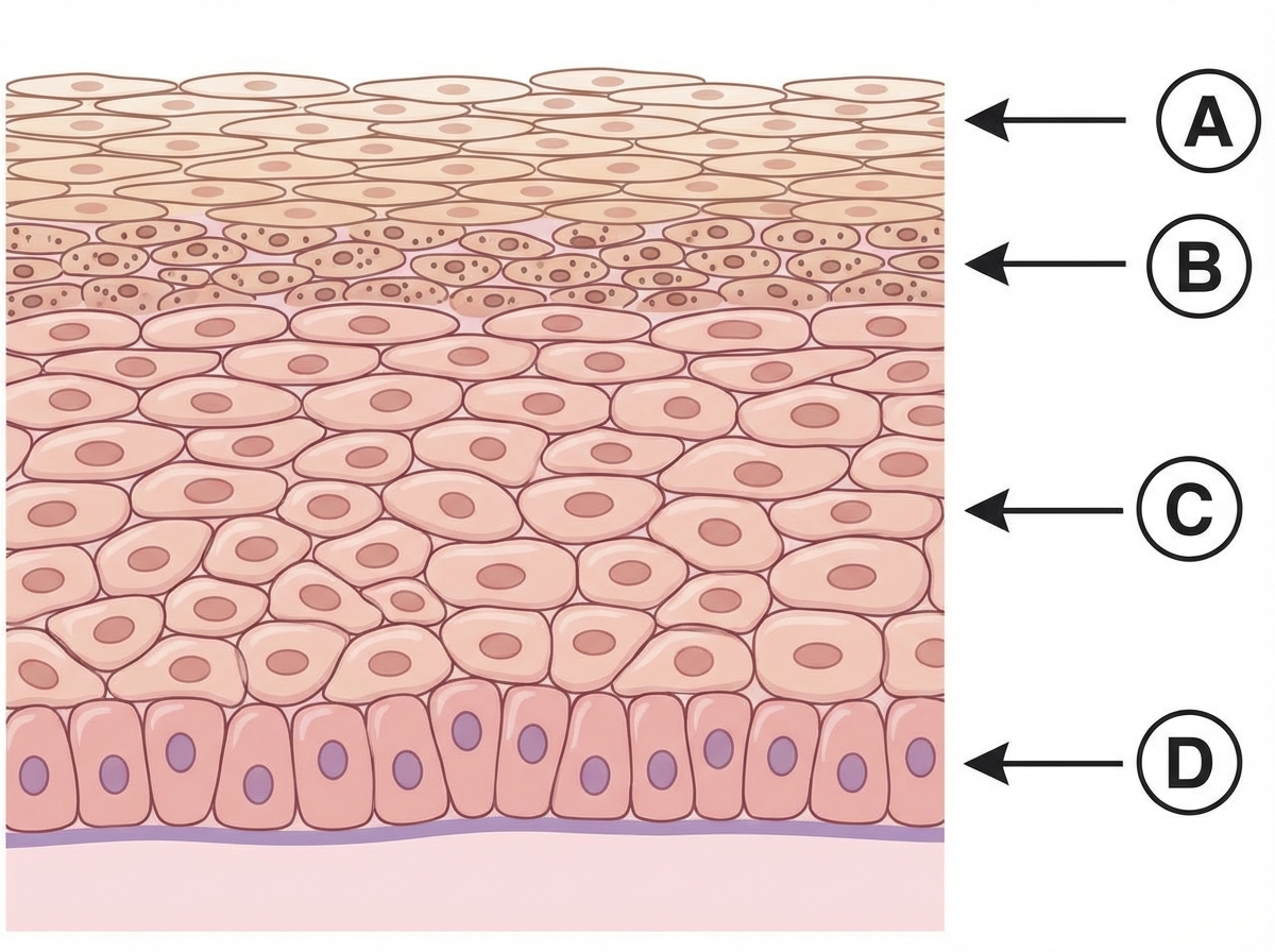
Q24
Which of the following layers contains Odland bodies?
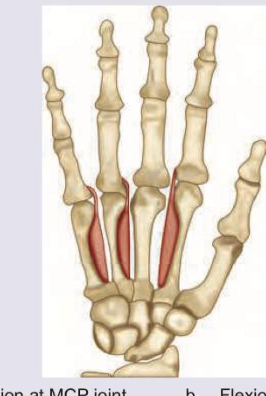
Q25
Identify the function of the muscles marked in red:
Q26
Which is correct about structures marked as "X" found in smooth muscle?

Q27
Which of the following has maximum smooth muscle as compared to wall thickness?