All SubjectsAnatomy (104)Anesthesiology (21)Biochemistry (179)Community Medicine (104)Dental (9)Dermatology (21)ENT (2)Forensic Medicine (41)General Medicine (2)Internal Medicine (79)Microbiology (83)Obstetrics and Gynecology (63)Ophthalmology (68)Orthopaedics (36)Pathology (82)Pediatrics (43)Pharmacology (85)Physiology (91)Psychiatry (2)Psychiatry (20)Radiology (28)Surgery (53)
Q11
Which of the following conditions characteristically causes bilateral hypertranslucency of lung fields on chest X-ray?
Q12
What is the most effective imaging method for the diagnosis of adenomyosis?
Q13
Investigation of choice for multiple sclerosis
Q14
Rat tail appearance in contrast radiography is seen in?
Q15
Which of the following appears the same on both T1 and T2 weighted MRI sequences?
Q16
Investigation of choice for screening of proximal internal carotid artery stenosis is :
Q17
Which of the following techniques uses piezoelectric crystals?
Q18
Which of the following conditions can cause periosteal reactions?
Q19
Rim sign in IVP is seen in
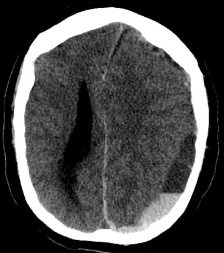
Q20
A polytrauma patient's CT brain shows a crescent-shaped extra-axial collection with a concave inner margin. What is the most likely diagnosis?