All (286)Anatomy (26)Anesthesiology (5)Biochemistry (20)Community Medicine (21)Dermatology (10)ENT (11)Forensic Medicine (9)General Medicine (1)Internal Medicine (33)Microbiology (14)Obstetrics and Gynecology (23)Ophthalmology (7)Orthopaedics (5)Pathology (15)Pediatrics (16)Pharmacology (17)Physiology (11)Psychiatry (6)Radiology (11)Surgery (25)
Q271
A patient presented with multiple painful blisters on an erythematous base along a dermatome on the trunk, as shown in the image. What is the diagnosis? 
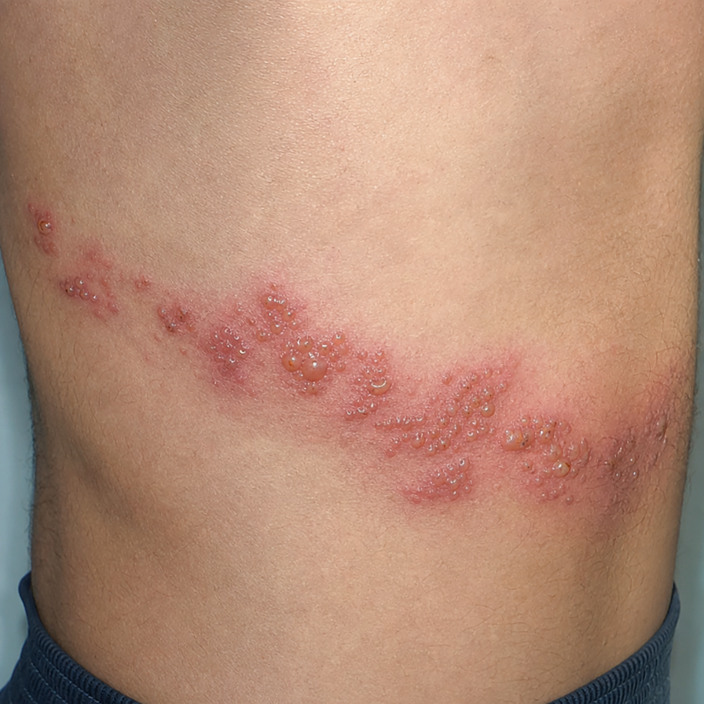
Q272
A patient presented with multiple painful blisters on an erythematous base along a dermatome on the trunk as shown in the image. What is the diagnosis?
Q273
A 45-year-old truck driver with a history of multiple sex partners presented to the dermatological department, as shown below. What is the likely diagnosis?