All (286)Anatomy (26)Anesthesiology (5)Biochemistry (20)Community Medicine (21)Dermatology (10)ENT (11)Forensic Medicine (9)General Medicine (1)Internal Medicine (33)Microbiology (14)Obstetrics and Gynecology (23)Ophthalmology (7)Orthopaedics (5)Pathology (15)Pediatrics (16)Pharmacology (17)Physiology (11)Psychiatry (6)Radiology (11)Surgery (25)
Q221
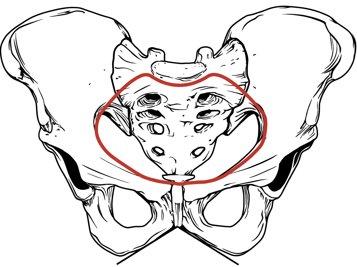
Identify the type of pelvis in the given image.