All (286)Anatomy (26)Anesthesiology (5)Biochemistry (20)Community Medicine (21)Dermatology (10)ENT (11)Forensic Medicine (9)General Medicine (1)Internal Medicine (33)Microbiology (14)Obstetrics and Gynecology (23)Ophthalmology (7)Orthopaedics (5)Pathology (15)Pediatrics (16)Pharmacology (17)Physiology (11)Psychiatry (6)Radiology (11)Surgery (25)
Q211
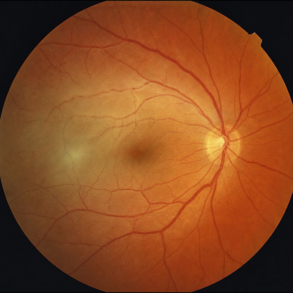
Fundoscopy findings are shown in the image below. What is the most likely diagnosis? 
Q212
Fundoscopy findings are shown in the image below. What is the most likely diagnosis?