All (286)Anatomy (26)Anesthesiology (5)Biochemistry (20)Community Medicine (21)Dermatology (10)ENT (11)Forensic Medicine (9)General Medicine (1)Internal Medicine (33)Microbiology (14)Obstetrics and Gynecology (23)Ophthalmology (7)Orthopaedics (5)Pathology (15)Pediatrics (16)Pharmacology (17)Physiology (11)Psychiatry (6)Radiology (11)Surgery (25)
Q201
In which of the following situations is ventouse not used?
Q202
Which of the following is not an indication for a cesarean section?
Q203
What is the level of the uterus immediately after delivery?
Q204
Which of the following procedures is done with the instrument shown below?
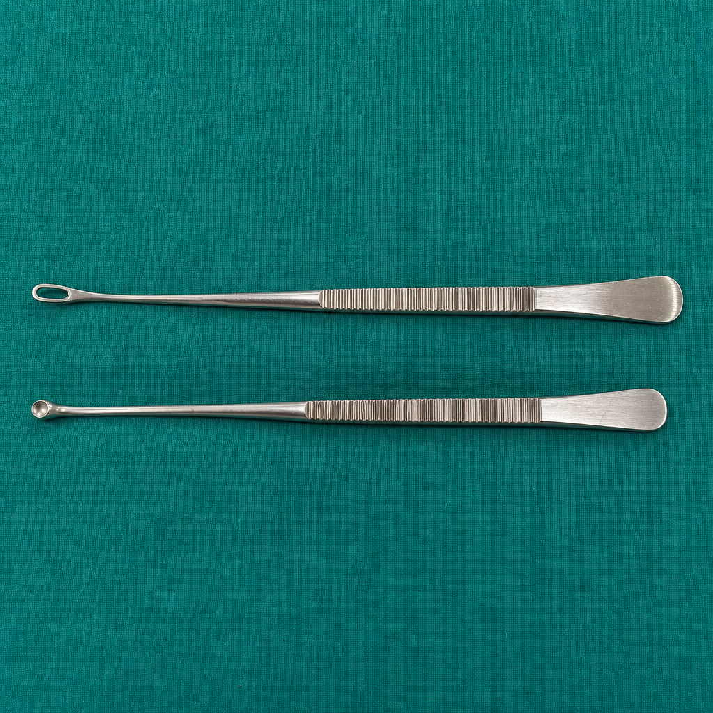
Q205
Which of the following procedures is done with the instrument shown below? 
Q206
Which of the following is the most common location of implantation in ectopic pregnancy? 