All SubjectsAnatomy (26)Anesthesiology (5)Biochemistry (20)Community Medicine (21)Dermatology (10)ENT (11)Forensic Medicine (9)General Medicine (1)Internal Medicine (33)Microbiology (14)Obstetrics and Gynecology (23)Ophthalmology (7)Orthopaedics (5)Pathology (15)Pediatrics (16)Pharmacology (17)Physiology (11)Psychiatry (6)Radiology (11)Surgery (25)
Q11
If the division in the zygote occurs between 9-12 days after fertilization, which of the following twins is expected?
Q12
Identify the segment marked in red in the image below.
Q13
Identify the rib highlighted in the X-ray.
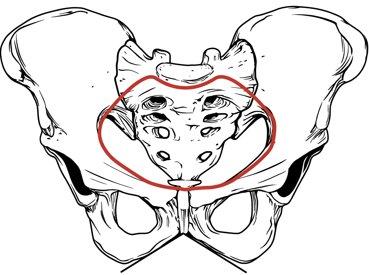
Q14
Identify the structure marked in the image given below.
Q15
Identify the type of pelvis in the given image.
Q16
Which of the following structures passes through the foramen marked by the arrow? 
Q17
Identify the nerve indicated by the arrow in the image.
Q18
Which nerve can be injured when a fracture is sustained in the area marked with the red arrow in the image? 
Q19
The condition seen in the image results from failure of fusion of:
Q20
Identify the given histology slide: