Molecular Imaging Indian Medical PG Practice Questions and MCQs
Practice Indian Medical PG questions for Molecular Imaging. These multiple choice questions (MCQs) cover important concepts and help you prepare for your exams.
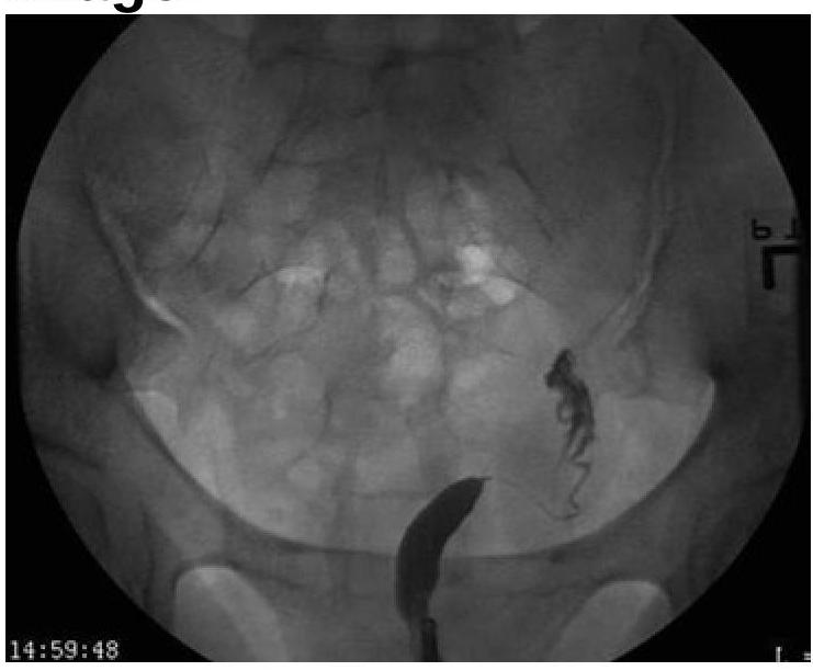
Molecular Imaging Indian Medical PG Question 1: Based on the provided image, which of the following is the correct diagnosis?
- A. Uterus didelphys
- B. Bicornuate Uterus
- C. Unicornuate Uterus (Correct Answer)
- D. Septate uterus
Molecular Imaging Explanation: ***Unicornuate Uterus***
- The image distinctly shows **only one fallopian tube and one rudimentary uterine horn** on the right side, indicating a unicornuate uterus.
- This malformation results from the **incomplete development of one Müllerian duct**, leading to a single, banana-shaped uterine cavity.
*Uterus didelphys*
- This condition involves **two completely separate uteri**, each with its own cervix and vagina.
- The image does not show evidence of two distinct uterine bodies or cervices.
*Bicornuate Uterus*
- A bicornuate uterus is characterized by **two uterine horns that fuse caudally**, creating a heart-shaped appearance with a shared cervix.
- The image clearly lacks the characteristic heart shape and shows only one functional horn.
*Septate uterus*
- A septate uterus has a **fibrous or muscular septum** dividing the uterine cavity, while the external uterine contour remains normal.
- The image does not show a septum or a normal external uterine contour with an internal division; instead, it presents with a single underdeveloped horn.
Molecular Imaging Indian Medical PG Question 2: Which of the following statements best describes the mechanism of action of insulin on target cells?
- A. Insulin binds to a receptor on the outer surface of the plasma membrane, activating adenylate cyclase through the Gs protein.
- B. Insulin binds to a cytoplasmic receptor and is transferred as a hormone receptor complex to the nucleus to modulate gene expression.
- C. Insulin enters the cell and causes the release of calcium ions from intracellular stores.
- D. Insulin binds to a transmembrane receptor on the outer surface of the plasma membrane, activating the tyrosine kinase in the cytosolic domain of the receptor. (Correct Answer)
Molecular Imaging Explanation: ***Insulin binds to a transmembrane receptor on the outer surface of the plasma membrane, activating the tyrosine kinase in the cytosolic domain of the receptor.***
- **Insulin** is a **peptide hormone** and cannot freely pass through the lipid bilayer, thus it binds to a **transmembrane receptor** on the cell surface.
- This binding leads to the activation of the receptor's intrinsic **tyrosine kinase activity** in the intracellular domain, initiating a signaling cascade.
*Insulin binds to a cytoplasmic receptor and is transferred as a hormone receptor complex to the nucleus to modulate gene expression.*
- This mechanism describes the action of **steroid hormones**, which are lipid-soluble and can cross the cell membrane, binding to **intracellular receptors**.
- **Insulin** acts via a **cell surface receptor** and its downstream effects are mediated through signal transduction pathways, not direct nuclear translocation.
*Insulin binds to a receptor on the outer surface of the plasma membrane, activating adenylate cyclase through the Gs protein.*
- This mechanism is characteristic of **G-protein coupled receptors (GPCRs)**, which activate or inhibit enzymes like adenylate cyclase via G-proteins to produce second messengers like cyclic AMP.
- The **insulin receptor** is a **receptor tyrosine kinase**, not a GPCR, and does not directly activate adenylate cyclase via Gs protein.
*Insulin enters the cell and causes the release of calcium ions from intracellular stores.*
- While some hormones and neurotransmitters can trigger the release of intracellular **calcium ions**, this is typically mediated by specific pathways (e.g., GPCRs linked to phospholipase C).
- **Insulin** does not directly enter target cells to cause calcium release; its actions are primarily mediated through receptor tyrosine kinase signaling pathways.
Molecular Imaging Indian Medical PG Question 3: Gold standard investigation for breast carcinoma screening in a patient with silicone breast implants
- A. Mammography
- B. CT scan
- C. USG
- D. MRI (Correct Answer)
Molecular Imaging Explanation: ***MRI***
- **MRI** is considered the **gold standard** for breast cancer screening in patients with silicone breast implants due to its superior ability to visualize breast tissue through the implant and detect subtle lesions.
- It offers **high sensitivity** in detecting both implant rupture and early malignancies, often providing better clarity than mammography in augmented breasts where implants can obscure tissue.
*Mammography*
- While a standard screening tool, **mammography** can be limited in patients with silicone implants because the implants can **obscure adjacent breast tissue**, making detection of small masses challenging.
- Special views (e.g., **Eklund views**) can be used, but sensitivity is still reduced compared to MRI in augmented breasts.
*CT scan*
- **CT scans** are not routinely used for primary breast cancer screening due to their use of **ionizing radiation** and lower sensitivity for detecting early breast lesions compared to MRI.
- CT is more commonly used for **staging** advanced cancers or evaluating complex masses detected by other modalities.
*USG*
- **Ultrasound (USG)** is a valuable complementary tool, especially for evaluating palpable lumps or clarifying findings from mammography, but it is **operator-dependent** and has a lower overall sensitivity for general screening compared to MRI.
- It is particularly useful for differentiating between **cystic and solid masses** and detecting implant ruptures but is not the gold standard for comprehensive screening in augmented breasts.
Molecular Imaging Indian Medical PG Question 4: Identify the gene commonly involved in the condition shown in the image?
- A. RAS
- B. RET
- C. BRAF V600E (Correct Answer)
- D. P53
Molecular Imaging Explanation: ***BRAF V600E***
- The image displays cells with **Langerhans cell morphology**, including folded nuclei and abundant pale cytoplasm, which are characteristic of **Langerhans cell histiocytosis (LCH)** [1].
- The **BRAF V600E mutation** is the most common genetic alteration found in LCH, present in about 50-60% of cases and activating the MAPK pathway [1].
*RAS*
- **RAS mutations** are frequently seen in various cancers, including colorectal adenocarcinoma, pancreatic adenocarcinoma, and non-small cell lung cancer.
- While RAS pathway activation can occur in LCH, a direct RAS mutation is not the most common genetic driver; rather, downstream effectors like BRAF V600E are more prominent [1].
*RET*
- **RET mutations** are primarily associated with **medullary thyroid carcinoma** (in both sporadic and inherited forms like MEN 2A and MEN 2B) and can also be found in certain types of lung cancer.
- They are not a characteristic genetic alteration for Langerhans cell histiocytosis.
*P53*
- The **TP53 gene** encodes the tumor suppressor protein p53, and mutations in this gene are among the most frequent genetic alterations across a wide spectrum of human cancers.
- Although p53 plays a critical role in cell cycle regulation and apoptosis, it is not a primary or common driver mutation specifically associated with Langerhans cell histiocytosis [1].
**References:**
[1] Kumar V, Abbas AK, et al.. Robbins and Cotran Pathologic Basis of Disease. 9th ed. Diseases of White Blood Cells, Lymph Nodes, Spleen, and Thymus, pp. 629-630.
Molecular Imaging Indian Medical PG Question 5: Investigation of choice for vascular ring around airway:
- A. PET
- B. Catheter directed angiography
- C. MRI
- D. CT (Correct Answer)
Molecular Imaging Explanation: ***CT***
- **CT angiography (CTA)** is the **investigation of choice** for diagnosing vascular rings due to its ability to provide detailed anatomical visualization of the great vessels and their relationship to the trachea and esophagus.
- It offers high spatial resolution, allowing precise identification of the type of vascular anomaly, the degree of **airway and esophageal compression**, and guiding surgical planning.
*PET*
- **PET scans** are primarily used for assessing **metabolic activity**, particularly in oncology or to evaluate organ function, and do not provide sufficient anatomical detail for vascular rings.
- While it can detect metabolically active lesions, it is **not suitable** for visualizing the structural abnormalities of blood vessels and their compressive effects on the airway.
*Catheter directed angiography*
- **Catheter-directed angiography** is an **invasive procedure** involving radiation and contrast, primarily used for assessing blood flow dynamics, identifying stenosis, or guiding interventions.
- While it can visualize vessels, CTA is **less invasive**, provides comparable or superior anatomical detail for vascular rings, and is generally preferred for initial diagnosis.
*MRI*
- **MRI** can provide good soft tissue contrast and visualize vascular structures without radiation, but it is often **less readily available** and can be more challenging for pediatric patients due to the need for sedation and longer scan times.
- For comprehensive anatomical detail including bone and calcifications, and in patients who might struggle with breath-holding, **CT angiography** often offers clearer and more consistent images of complex vascular anatomy.
More Molecular Imaging Indian Medical PG questions available in the OnCourse app. Practice MCQs, flashcards, and get detailed explanations.