Contrast and Radiological Procedures Indian Medical PG Practice Questions and MCQs
Practice Indian Medical PG questions for Contrast and Radiological Procedures. These multiple choice questions (MCQs) cover important concepts and help you prepare for your exams.
Contrast and Radiological Procedures Indian Medical PG Question 1: All of the following are advantages of the paralleling technique except?
- A. An excellent bone level assessment
- B. The shadow of the zygomatic bone frequently overlies the roots of the upper molars (Correct Answer)
- C. No elongation or foreshortening seen in the periapical region
- D. Interproximal caries is clearly indicated
Contrast and Radiological Procedures Explanation: **Explanation:**
The **Paralleling Technique** (also known as the Extension Cone Paralleling or Right-Angle technique) is the gold standard for intraoral periapical radiographs (IOPA). It involves placing the film/sensor parallel to the long axis of the tooth, with the X-ray beam directed perpendicularly to both.
**Why Option B is the Correct Answer (The "Except"):**
In the paralleling technique, the film is placed further away from the teeth to achieve parallelism. This positioning, combined with a perpendicular beam, ensures that the **zygomatic bone (malar process) is projected superiorly**, away from the roots of the maxillary molars. In contrast, the *Bisecting Angle Technique* often causes the zygomatic bone to be superimposed over the molar roots due to the steep vertical angulation required. Therefore, avoiding this shadow is an **advantage** of the paralleling technique, making the statement in Option B a disadvantage/limitation that does not apply here.
**Analysis of Incorrect Options:**
* **Option A:** Because the beam is perpendicular to the tooth and film, there is minimal distortion, allowing for an **accurate assessment of alveolar bone levels**, crucial for periodontology.
* **Option C:** The geometric accuracy of this technique prevents **elongation or foreshortening**, which are common errors in the bisecting angle technique.
* **Option D:** Since the beam passes directly through the contact points, **interproximal caries** are visualized with high clarity and minimal overlapping.
**Clinical Pearls for NEET-PG:**
* **Rule of Isometry:** This is the basis for the *Bisecting Angle Technique*, not the paralleling technique.
* **Increased Object-Film Distance:** A drawback of the paralleling technique is increased magnification, which is compensated for by using a **Long Cone (16 inches)** to ensure the X-rays are more parallel.
* **Patient Comfort:** The paralleling technique is often more difficult to perform in patients with a shallow palate or small mouth.
Contrast and Radiological Procedures Indian Medical PG Question 2: In which of the following conditions is ground glass appearance of the maxillary sinus seen?
- A. Maxillary sinusitis
- B. Maxillary carcinoma
- C. Maxillary polyp
- D. Maxillary fibrous dysplasia (Correct Answer)
Contrast and Radiological Procedures Explanation: **Explanation:**
The "ground glass" appearance is a classic radiological hallmark of **Fibrous Dysplasia**. This condition occurs due to the replacement of normal medullary bone with cellular fibrous tissue and irregular bony trabeculae (woven bone). On imaging (X-ray or CT), this disorganized mineralization results in a characteristic smoky, hazy, or "ground glass" opacity that lacks a distinct cortical-medullary margin. When it involves the facial bones (craniofacial fibrous dysplasia), the maxillary sinus is frequently affected, appearing opacified with a dense, frosted-glass texture.
**Analysis of Incorrect Options:**
* **Maxillary Sinusitis (A):** Typically presents as mucosal thickening or an air-fluid level. On imaging, it appears as a simple opacification (radio-opacity) rather than a textured ground-glass pattern.
* **Maxillary Carcinoma (B):** Usually presents as a soft tissue mass causing **bone destruction** and aggressive erosion of the sinus walls. It does not produce the characteristic organized hazy mineralization of fibrous dysplasia.
* **Maxillary Polyp (C):** Appears as a smooth, rounded, soft-tissue density within the sinus. It may cause expansion if large, but the internal density is that of soft tissue/fluid, not bone.
**High-Yield Clinical Pearls for NEET-PG:**
* **Fibrous Dysplasia:** Look for the "Ground Glass" appearance on CT. It can be Monostotic (one bone) or Polyostotic (multiple bones).
* **McCune-Albright Syndrome:** Triad of Polyostotic fibrous dysplasia, Café-au-lait spots (Coast of Maine borders), and Precocious puberty.
* **Lichtenstein-Jaffe Syndrome:** Polyostotic fibrous dysplasia with Café-au-lait spots but *without* endocrine involvement.
* **Cherubism:** A related condition involving bilateral, symmetrical multilocular cystic expansion of the jaws (soap-bubble appearance).
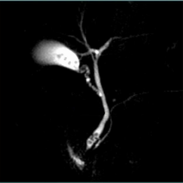
Contrast and Radiological Procedures Indian Medical PG Question 3: What radiological finding is described as a "coiled spring appearance"?
- A. Intussusception (Correct Answer)
- B. Achalasia
- C. Duodenal perforation
- D. Chronic pancreatitis
Contrast and Radiological Procedures Explanation: ### Explanation
**Correct Option: A. Intussusception**
The "coiled spring appearance" is a classic radiological sign of **intussusception**, most commonly seen during a **Barium Enema** or air contrast enema. It occurs when the invaginating portion of the bowel (intussusceptum) is surrounded by the receiving portion (intussuscipiens). The contrast material gets trapped in the thin space between these two layers, outlining the mucosal folds and creating a striated, spring-like appearance. On **Ultrasound**, this same pathology presents as the "Target sign" or "Donut sign" in cross-section and the "Pseudokidney sign" in longitudinal section.
**Incorrect Options:**
* **B. Achalasia:** Characterized by a **"Bird’s beak"** or "Rat-tail" appearance on Barium Swallow due to the failure of the lower esophageal sphincter to relax.
* **C. Duodenal perforation:** Typically presents as **"Gas under the diaphragm"** (Pneumoperitoneum) on an erect X-ray abdomen.
* **D. Chronic pancreatitis:** Classically shows **diffuse pancreatic calcifications** on X-ray or CT, and a "Chain of lakes" appearance (dilated, irregular pancreatic duct) on MRCP/ERCP.
**High-Yield Clinical Pearls for NEET-PG:**
* **Clinical Triad of Intussusception:** Intermittent abdominal pain, palpable sausage-shaped mass (usually in the right upper quadrant), and **"Red currant jelly" stools**.
* **Dance’s Sign:** An empty right iliac fossa due to the migration of the cecum into the hepatic flexure.
* **Management:** Hydrostatic or pneumatic reduction is the first-line treatment in stable pediatric cases; surgery is indicated if there are signs of peritonitis or gangrene.
Contrast and Radiological Procedures Indian Medical PG Question 4: What is the most common source of error leading to a false positive finding of dental caries?
- A. Cervical burnout (Correct Answer)
- B. Fluorosis
- C. Dental pigmentation
- D. All of the above
Contrast and Radiological Procedures Explanation: **Explanation:**
**Cervical burnout** is the most common radiographic artifact mimicking dental caries. It appears as a radiolucent (dark) area at the neck of the tooth, between the enamel cap and the alveolar bone crest.
**Why it occurs:** This phenomenon is due to the **anatomical configuration** of the tooth. At the cervical region, there is a relative lack of tooth mass compared to the crown (protected by thick enamel) and the root (surrounded by bone). Because fewer X-rays are absorbed in this narrow neck area, more radiation reaches the film, creating a radiolucency that clinicians often mistake for proximal or root caries.
**Analysis of Incorrect Options:**
* **Fluorosis (B):** This is a developmental disturbance caused by excess fluoride. While it causes physical changes like mottling or pitting of the enamel, it does not typically create localized radiolucencies on a radiograph that mimic the specific appearance of caries.
* **Dental Pigmentation (C):** Surface staining or pigmentation is a clinical visual finding. Since these pigments do not significantly alter the density of the tooth structure, they do not produce false-positive radiolucencies on an X-ray.
**High-Yield Clinical Pearls for NEET-PG:**
* **Differential Diagnosis:** To distinguish cervical burnout from true caries, look for the **intactness of the tooth outline**. Burnout disappears when the X-ray angle is changed, whereas true caries remains visible.
* **Mach Band Effect:** Another common optical illusion in radiology where the high contrast between enamel and dentin creates a perceived dark line, often leading to a false diagnosis of "occlusal caries."
* **Adumbration:** This is the technical term for the shadowing effect seen in cervical burnout.
Contrast and Radiological Procedures Indian Medical PG Question 5: What is the imaging of choice for urethral trauma?
- A. Ascending urethrogram (Correct Answer)
- B. Descending urethrogram
- C. Ultrasound (USG)
- D. CT scan
Contrast and Radiological Procedures Explanation: **Explanation:**
The imaging of choice for suspected urethral trauma is an **Ascending Urethrogram (RGU - Retrograde Urethrogram)**.
**Why Ascending Urethrogram is Correct:**
In cases of suspected urethral injury (often indicated by clinical signs like blood at the meatus, high-riding prostate, or inability to void), the primary goal is to assess the integrity of the urethral lumen. RGU involves the retrograde injection of water-soluble contrast into the external meatus. It is the most sensitive and specific test for identifying the **site, nature, and extent of a urethral tear** (partial vs. complete) before any attempt at catheterization, which could convert a partial tear into a complete one.
**Why Other Options are Incorrect:**
* **Descending Urethrogram (MCU/VCUG):** This requires the bladder to be full of contrast, usually via a suprapubic catheter or by waiting for excreted IV contrast. It is better for evaluating the posterior urethra during voiding but is not the initial investigation for acute trauma.
* **Ultrasound (USG):** While useful for evaluating the bladder or scrotal hematomas, USG lacks the resolution to accurately map urethral mucosal disruptions or extravasation in an acute setting.
* **CT Scan:** CT is the gold standard for evaluating stable blunt abdominal trauma and pelvic fractures, but it is insensitive for identifying specific urethral mucosal injuries.
**Clinical Pearls for NEET-PG:**
* **Classic Triad of Urethral Injury:** Blood at the meatus, inability to void, and a palpable distended bladder.
* **Membranous Urethra:** The most common site of injury in pelvic fractures (Posterior Urethra).
* **Bulbar Urethra:** The most common site of injury in "straddle" injuries (Anterior Urethra).
* **Contraindication:** Never perform a blind Foley catheterization if urethral trauma is suspected; perform an RGU first.
More Contrast and Radiological Procedures Indian Medical PG questions available in the OnCourse app. Practice MCQs, flashcards, and get detailed explanations.