Pediatric Dermatology Indian Medical PG Practice Questions and MCQs
Practice Indian Medical PG questions for Pediatric Dermatology. These multiple choice questions (MCQs) cover important concepts and help you prepare for your exams.
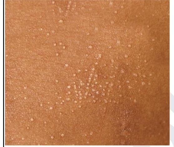
Pediatric Dermatology Indian Medical PG Question 1: A child presented with asymptomatic lesions on the forearm and on the shaft of the penis. The lesions on the forearm are shown below. What is the most likely diagnosis?
- A. Lichen planus
- B. Lichen nitidus (Correct Answer)
- C. Scabies
- D. Scrofuloderma
Pediatric Dermatology Explanation: ***Lichen nitidus***
- Presents as **multiple, asymptomatic, tiny (1-2 mm), shiny, dome-shaped papules** that are often skin-colored or slightly hypopigmented, as seen in the image and described.
- Common sites include the **forearms, penis, abdomen, and flexural areas**, consistent with the case presentation.
*Lichen planus*
- Characterized by **purplish, polygonal, planar, pruritic papules and plaques**, often with **Wickham's striae**, which are not seen in the image.
- While it can affect the penis, its lesions are typically more intensely colored and often symptomatic (**itchy**), unlike the asymptomatic lesions described.
*Scabies*
- Presents with intensely **pruritic papules, vesicles, and burrows**, especially in the web spaces of fingers, wrists, axillae, and genitalia, which are very symptomatic and not usually described as shiny papules.
- The primary symptom is **severe itching**, which is absent in this patient.
*Scrofuloderma*
- A form of **cutaneous tuberculosis** presenting as cold abscesses that eventually rupture to form ulcers, sinuses, and scars.
- The image shows distinct, small papules, not ulcerating or scarring lesions characteristic of scrofuloderma.
Pediatric Dermatology Indian Medical PG Question 2: A child has a rash. His family history is positive for asthma. What could be the most probable diagnosis?
- A. Seborrheic dermatitis
- B. Atopic dermatitis (Correct Answer)
- C. Allergic contact dermatitis
- D. Erysipelas
Pediatric Dermatology Explanation: ***Atopic dermatitis***
- The presence of a rash in a child with a family history of **asthma** strongly suggests atopic dermatitis, as it is part of the **atopic triad** (eczema, asthma, allergic rhinitis).
- Atopic dermatitis often presents with **erythematous, pruritic patches** and plaques, commonly affecting flexural areas like the antecubital and popliteal fossae, as well as the face and neck in younger children.
*Seborrheic dermatitis*
- This condition typically presents with **greasy, yellowish scales** on an erythematous base, often affecting areas rich in sebaceous glands such as the scalp, face (nasolabial folds), and chest.
- While it can occur in infants, it does not have the strong association with a family history of asthma seen in atopic dermatitis.
*Allergic contact dermatitis*
- This rash results from an **exposure to an allergen**, leading to a localized, erythematous, and pruritic eruption, often with vesicles or bullae, at the site of contact.
- The history does not provide information about a specific allergen exposure, and while it could produce a similar-looking rash, the family history of asthma points more strongly to atopic diathesis.
*Erysipelas*
- Erysipelas is a superficial skin infection, usually caused by *Streptococcus pyogenes*, presenting as a **well-demarcated, intensely erythematous, warm, and painful rash** with a raised border.
- This is an **acute bacterial infection** and would typically be accompanied by systemic symptoms like fever and chills, which are not mentioned in the child's presentation.
Pediatric Dermatology Indian Medical PG Question 3: A child with fever for 6 days, strawberry tongue, conjunctival congestion with peeling of skin. What will be the treatment option for this child?
- A. Antibiotics
- B. Steroids
- C. Antipyretics
- D. IVIG (Correct Answer)
Pediatric Dermatology Explanation: ***IVIG***
- The constellation of **fever for 6 days (prolonged fever)**, **strawberry tongue**, **conjunctival congestion**, and **peeling skin** is highly indicative of **Kawasaki disease**.
- **Intravenous immunoglobulin (IVIG) 2 g/kg as a single infusion** is the cornerstone of treatment for Kawasaki disease to reduce the risk of **coronary artery aneurysms** (from ~25% to <5%).
- IVIG should be administered within **10 days of fever onset** for maximum efficacy.
- **High-dose aspirin** (80-100 mg/kg/day) is given concurrently until the fever subsides, then switched to low-dose aspirin (3-5 mg/kg/day) for antiplatelet effect.
*Antibiotics*
- Kawasaki disease is a **vasculitis**, not a bacterial infection, so antibiotics are ineffective.
- While other conditions like scarlet fever can present with strawberry tongue, the prolonged fever and other classic Kawasaki features differentiate it.
*Steroids*
- While steroids can reduce inflammation, they are **not the primary treatment** for Kawasaki disease and are typically used in conjunction with IVIG in **refractory cases** or for IVIG-resistant disease.
- **Monotherapy with steroids** is not recommended for acute Kawasaki disease due to potential for increased aneurysm risk.
*Antipyretics*
- **Antipyretics** like acetaminophen can help manage the fever symptomatically.
- However, they **do not treat the underlying vasculitis** or prevent the serious cardiac complications of Kawasaki disease.
- Note: **NSAIDs like ibuprofen should be avoided** when high-dose aspirin is being used due to risk of drug interactions.
Pediatric Dermatology Indian Medical PG Question 4: Match the following scale types with their lesions.
| Scales | Lesions |
| :-- | :-- |
| 1. Collarette scales | a. Pityriasis versicolour |
| 2. Silvery scales | b. Pityriasis rosea |
| 3. Mica-like scales | c. Psoriasis |
| 4. Branny scales | d. Pityriasis lichenoides |
- A. 1-d, 2-c, 3-a, 4-b
- B. 1-c, 2-b, 3-d, 4-a
- C. 1-a, 2-b, 3-d, 4-c
- D. 1-b, 2-c, 3-d, 4-a (Correct Answer)
Pediatric Dermatology Explanation: ***1-b, 2-c, 3-d, 4-a***
- **Collarette scales** are pathognomonic of **Pityriasis rosea**, appearing as fine, trailing scales around the periphery of oval lesions in a "Christmas tree" distribution.
- **Silvery scales** are the classic hallmark of **Psoriasis**, presenting as thick, adherent, silvery-white scales overlying well-demarcated erythematous plaques.
- **Mica-like scales** are characteristic of **Pityriasis lichenoides**, appearing as thick, shiny, adherent scales that can be peeled off like mica sheets.
- **Branny scales** are typical of **Pityriasis versicolor**, presenting as fine, powdery scales caused by **Malassezia** yeast overgrowth.
*1-d, 2-c, 3-a, 4-b*
- Incorrectly matches **collarette scales with Pityriasis lichenoides**, which typically presents with mica-like scales, not collarette scales.
- Misassociates **mica-like scales with Pityriasis versicolor**, which characteristically has branny (fine, powdery) scales.
*1-c, 2-b, 3-d, 4-a*
- Wrongly pairs **collarette scales with Psoriasis**, which is known for thick silvery scales, not peripheral collarette scales.
- Incorrectly matches **silvery scales with Pityriasis rosea**, which has collarette scales at lesion periphery, not silvery scales.
*1-a, 2-b, 3-d, 4-c*
- Falsely associates **collarette scales with Pityriasis versicolor**, which has branny scales from yeast infection, not collarette scales.
- Mismatches **branny scales with Psoriasis**, which has characteristic thick silvery scales, not fine powdery scales.
Pediatric Dermatology Indian Medical PG Question 5: A 15cm hyperpigmented macule on an adolescent male undergoes changes such as coarseness, growth of hair & acne. Diagnosis is?
- A. Melanocytic nevus
- B. Becker nevus (Correct Answer)
- C. Sebaceous nevus
- D. Sebaceous adenoma
Pediatric Dermatology Explanation: ***Becker nevus***
- A Becker nevus is a **hyperpigmented patch** that typically appears during adolescence in males, often on the shoulder or upper trunk.
- It characteristically becomes **hairy (hypertrichosis)**, more coarse, and can develop acne within the lesion, particularly during puberty due to androgen sensitivity.
*Melanocytic nevus*
- While melanocytic nevi are hyperpigmented, they generally do not show the characteristic changes of **coarseness, significant hair growth, or acne** within the lesion during adolescence.
- They are typically stable in size and texture after initial development, with changes raising concern for **melanoma**.
*Sebaceous nevus*
- A sebaceous nevus is a **congenital lesion** often appearing as a yellowish-orange, waxy, or bumpy patch, usually on the scalp or face.
- It does not typically present as a large, flat hyperpigmented macule that develops hair and acne in adolescence; instead, it may become verrucous or develop tumors in adulthood.
*Sebaceous adenoma*
- A sebaceous adenoma is a **benign tumor** of the sebaceous glands, usually appearing as a small, solitary, flesh-colored to yellowish papule or nodule, especially on the face.
- It is not typically seen as a large, hyperpigmented macule that grows hair and acne over a broad area, as described in the question.
More Pediatric Dermatology Indian Medical PG questions available in the OnCourse app. Practice MCQs, flashcards, and get detailed explanations.