Surface and Radiological Anatomy Indian Medical PG Practice Questions and MCQs
Practice Indian Medical PG questions for Surface and Radiological Anatomy. These multiple choice questions (MCQs) cover important concepts and help you prepare for your exams.
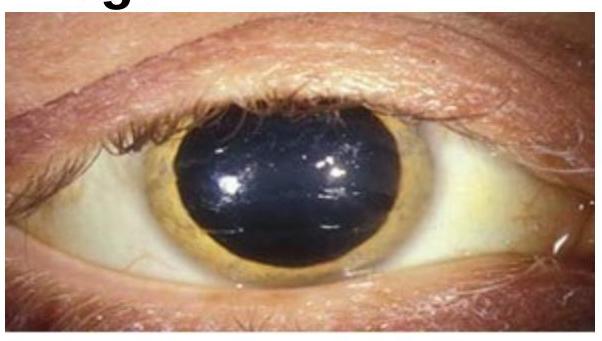
Surface and Radiological Anatomy Indian Medical PG Question 1: Choose the best method of diagnosis for the clinical sign represented in the image.
- A. Serum copper
- B. Serum ceruloplasmin (Correct Answer)
- C. Karyotyping
- D. PCR
Surface and Radiological Anatomy Explanation: ***Serum ceruloplasmin***
- The image shows a **Kayser-Fleischer ring**, a greenish-brown discoloration in the periphery of the cornea, which is pathognomonic for **Wilson's disease**.
- **Wilson's disease** is a genetic disorder of copper metabolism characterized by **low serum ceruloplasmin** levels (the primary copper-carrying protein in the blood) and increased copper deposition in various tissues.
*Serum copper*
- While Wilson's disease involves copper accumulation, **total serum copper** can be normal or even elevated due to widespread tissue damage releasing copper into the circulation, making it an unreliable diagnostic marker on its own.
- A low serum copper level can be seen, but it is not as specific as low ceruloplasmin, as much of the copper in serum is bound to ceruloplasmin.
*Karyotyping*
- **Karyotyping** is used to analyze the number and structure of chromosomes and is primarily indicated for diagnosing chromosomal abnormalities, such as Down syndrome or Turner syndrome.
- It is not relevant for diagnosing metabolic disorders like Wilson's disease, which is caused by a mutation in a single gene (ATP7B), not a chromosomal aberration.
*PCR*
- **PCR (Polymerase Chain Reaction)** is a technique used to amplify DNA sequences and can be used for genetic testing to identify specific mutations.
- While genetic testing for the **ATP7B gene** mutation is a confirmatory test for Wilson's disease, it is not the primary or best method for initial diagnosis, especially when classic clinical signs and biochemical markers (like low ceruloplasmin) are present.
Surface and Radiological Anatomy Indian Medical PG Question 2: Which of the following statements regarding lymphoedema are correct?
1. Patients experience constant dull ache and even severe pain sometimes
2. Manual lymphatic drainage has a role
3. Primary lymphoedema is caused by congenital lymphatic dysplasia
4. Nonne Milroy's disease is a type of primary lymphoedema
Select the correct answer using the code given below:
- A. 1 and 2 only
- B. 1, 2, 3 and 4 (Correct Answer)
- C. 3 and 4 only
- D. 1, 2 and 3 only
Surface and Radiological Anatomy Explanation: ***1, 2, 3 and 4***
- All four statements are correct regarding lymphoedema. Patients often experience **constant dull ache and severe pain** due to the swelling and tissue changes.
- **Manual lymphatic drainage (MLD)** is a key component of complete decongestive therapy for lymphoedema, aiming to reduce swelling and improve lymphatic flow. **Primary lymphoedema** is indeed caused by **congenital lymphatic dysplasia**, which refers to abnormalities in lymphatic system development from birth. **Milroy's disease** (also known as Nonne-Milroy disease) is a specific type of primary lymphoedema characterized by early-onset lymphatic dysfunction.
*1 and 2 only*
- This option is incomplete as statements 3 and 4 are also correct.
- It correctly identifies the role of manual lymphatic drainage and the presence of pain in lymphoedema but omits other accurate facts.
*3 and 4 only*
- This option is incomplete as statements 1 and 2 are also correct.
- While correctly identifying the nature of primary lymphoedema and Milroy's disease, it misses other important aspects of lymphoedema.
*1, 2, and 3 only*
- This option is incomplete because statement 4, concerning Milroy's disease as a type of primary lymphoedema, is also correct.
- It provides correct information about pain, MLD, and the cause of primary lymphoedema but omits a specific example of primary lymphoedema.
Surface and Radiological Anatomy Indian Medical PG Question 3: A 50-year-old male presents with sharp, localized chest pain, worse with deep breaths and relieved by sitting up. ECG is normal. What is the most likely diagnosis?
- A. Pneumothorax
- B. Myocardial infarction
- C. Pleuritis
- D. Pericarditis (Correct Answer)
Surface and Radiological Anatomy Explanation: ***Pericarditis***
- The classic presentation of **sharp, localized chest pain** that is **worse with deep breaths** and **relieved by sitting up and leaning forward** is highly characteristic of pericarditis [2].
- A **normal ECG** makes other cardiac causes less likely, supporting the diagnosis of pericarditis, which can have diffuse ST elevation or PR depression as ECG findings, but a normal ECG doesn't rule it out, especially early on [2].
*Pneumothorax*
- While pneumothorax can cause **sharp chest pain** and be **respiratory variation**, it is typically associated with **dyspnea** and **diminished breath sounds** on examination, which are not mentioned here.
- The pain relief with sitting up is not characteristic of pneumothorax.
*Myocardial infarction*
- **Myocardial infarction** pain is typically described as a **heavy, pressure-like sensation**, often radiating to the arm, jaw, or back, and is usually not relieved by position changes [1].
- A **normal ECG** makes acute myocardial infarction less likely, though it does not entirely rule out non-ST elevation myocardial infarction (NSTEMI).
*Pleuritis*
- **Pleuritis** also causes **sharp, pleuritic chest pain** that worsens with deep inspiration or coughing.
- However, the classic relief with **sitting up and leaning forward** is more specific to pericarditis than pleuritis.
Surface and Radiological Anatomy Indian Medical PG Question 4: All cartilage is covered by perichondrium, except
- A. Hyaline
- B. Elastic
- C. Fibrocartilage (Correct Answer)
- D. All types of cartilage are covered by perichondrium.
Surface and Radiological Anatomy Explanation: ***Fibrocartilage***
- **Fibrocartilage** completely lacks a perichondrium as a defining characteristic of this cartilage type.
- This type of cartilage is found in structures like **intervertebral discs**, **pubic symphysis**, and **menisci**, where it provides strong support and shock absorption.
- It merges imperceptibly with surrounding dense connective tissue without a distinct perichondrial covering.
- Among all cartilage types, **fibrocartilage is the only type that NEVER has perichondrium**.
*Hyaline*
- Most **hyaline cartilage** (such as in the **trachea, bronchi, nose, larynx, and ribs**) is covered by a **perichondrium** that provides growth and nutrition.
- **Important exception:** **Articular cartilage** (covering joint surfaces) lacks perichondrium because it receives nutrition from synovial fluid, but this is a specific exception within the hyaline category [1].
- Since hyaline cartilage CAN have perichondrium in most locations, it is not the best answer to this "except" question.
*Elastic*
- **Elastic cartilage**, found in structures such as the **external ear (auricle)** and **epiglottis**, is always surrounded by a **perichondrium**.
- The perichondrium supports growth, provides nutrition, and aids in repair after injury.
*All types of cartilage are covered by perichondrium*
- This statement is **incorrect** because fibrocartilage never has a perichondrium.
- Additionally, articular hyaline cartilage also lacks perichondrium, making this statement doubly false [1].
Surface and Radiological Anatomy Indian Medical PG Question 5: Highest point of iliac crest is seen at?
- A. L3
- B. L4 (Correct Answer)
- C. S2
- D. S1
Surface and Radiological Anatomy Explanation: ***L4***
- The **highest point of the iliac crest** typically corresponds to the level of the **L4 vertebral body**.
- This anatomical landmark is crucial for procedures like **lumbar punctures** and determining the location for **epidural anesthesia**.
*L3*
- The L3 vertebral level is generally located slightly **above the highest point of the iliac crest**.
- While close, it is not the most consistent anatomical correlation for the highest point.
*S2*
- The **S2 vertebral level** is significantly **below the iliac crests**, marking the approximate midpoint of the sacroiliac joint.
- This level is used as a landmark for the **dermatome of the posterior thigh**.
*S1*
- The **S1 vertebral level** is also located **below the iliac crests**, forming the most superior segment of the sacrum.
- It is used as a landmark for the **dermatome of the lateral foot and posterior leg**, and corresponds to the ankle jerk reflex.
More Surface and Radiological Anatomy Indian Medical PG questions available in the OnCourse app. Practice MCQs, flashcards, and get detailed explanations.