Histology Indian Medical PG Practice Questions and MCQs
Practice Indian Medical PG questions for Histology. These multiple choice questions (MCQs) cover important concepts and help you prepare for your exams.
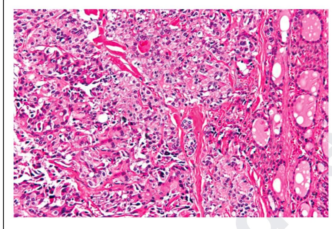
Histology Indian Medical PG Question 1: The following is a histopathological image of thyroid pathology. What is the diagnosis?
- A. Papillary carcinoma of thyroid
- B. Medullary carcinoma of thyroid (Correct Answer)
- C. Follicular carcinoma of thyroid
- D. Anaplastic carcinoma of thyroid
Histology Explanation: ***Medullary carcinoma of thyroid***
- This image shows sheets and nests of **polygonal to spindle-shaped cells**, which are characteristic of medullary thyroid carcinoma, especially when mixed with an **amyloid stroma** (seen as amorphous eosinophilic material) [2].
- The presence of **neuroendocrine features** and the production of **calcitonin** are hallmarks of these C-cell tumors [1], [2].
*Papillary carcinoma of thyroid*
- Characterized by **papillary architecture**, **ground-glass (Orphan Annie eye) nuclei**, nuclear grooves, and intranuclear cytoplasmic inclusions.
- These features are not prominently seen in the provided image.
*Follicular carcinoma of thyroid*
- Defined by an invasive growth pattern of **well-differentiated follicular cells** forming follicles, with either capsular or vascular invasion [2].
- The image does not show classic follicular architectural patterns or clear evidence of invasion in the absence of a capsule.
*Anaplastic carcinoma of thyroid*
- This is a highly aggressive and undifferentiated tumor with **marked pleomorphism**, bizarre giant cells, and high mitotic activity [2].
- While there is some pleomorphism, the overall pattern and cellular morphology in the image are more consistent with medullary carcinoma than the extreme anaplasia.
**References:**
[1] Kumar V, Abbas AK, et al.. Robbins and Cotran Pathologic Basis of Disease. 9th ed. The Endocrine System, pp. 1102-1103.
[2] Cross SS. Underwood's Pathology: A Clinical Approach. 6th ed. Common Clinical Problems From Liver And Biliary System Disease, pp. 428-431.
Histology Indian Medical PG Question 2: Arrange the following parts of sarcomere from periphery to center.
1. Z line
2. M line
3. A band
4. H zone
- A. 2,3,4,1
- B. 4,2,3,1
- C. 3,1,4,2
- D. 1,3,4,2 (Correct Answer)
Histology Explanation: ***1,3,4,2***
- The **Z line** is found at the **periphery** of the sarcomere, defining its boundaries and anchoring the **actin filaments**.
- Moving inwards, the **A band** is next, representing the entire length of the **myosin filament**, which may also overlap with actin.
- The **H zone** is located within the A band, comprising only **myosin filaments** without actin overlap.
- Finally, the **M line** is at the **center** of the sarcomere, bisecting the H zone and anchoring the myosin filaments.
*2,3,4,1*
- This sequence is incorrect because the **M line** is at the **center** and the **Z line** is at the **periphery**, which is the reverse of the expected order for from periphery to center.
- Such an arrangement would place the innermost structure first and outermost last, not reflecting the correct spatial organisation.
*4,2,3,1*
- This order is incorrect as the **H zone** and **M line** are more central, while the **Z line** is peripheral.
- Placing structures like the H zone and M line at the beginning does not align with arrangement from periphery to center.
*3,1,4,2*
- This option is incorrect because the **A band** includes both actin and myosin filaments, while the **Z line** is at the periphery of the sarcomere.
- The given order does not represent a progression from the periphery to the center of the sarcomere.
Histology Indian Medical PG Question 3: Demyelination is the major feature of Multiple Sclerosis. Which of the following cells forms myelin in the central nervous system?
- A. Astrocytes
- B. Ependymal cells
- C. Microglia
- D. Oligodendrocytes (Correct Answer)
Histology Explanation: ***Oligodendrocytes***
- **Oligodendrocytes** are glial cells exclusively found in the **central nervous system (CNS)** that produce and maintain the **myelin sheath**.
- The myelin sheath, formed by these cells, insulates axons and allows for rapid, efficient **saltatory conduction** of action potentials.
*Astrocytes*
- **Astrocytes** are star-shaped glial cells that provide structural and metabolic support for neurons, regulate the **blood-brain barrier**, and maintain the extracellular environment.
- They do not form myelin; instead, they play roles in **neurotransmission** and response to injury.
*Ependymal cells*
- **Ependymal cells** line the ventricles of the brain and the central canal of the spinal cord, forming an interface between the neural tissue and **cerebrospinal fluid (CSF)**.
- They are involved in **CSF production** and circulation, having no role in myelination.
*Microglia*
- **Microglia** are the resident immune cells of the CNS, functioning as **macrophages** to remove cellular debris, pathogens, and damaged neurons.
- They are crucial for immune surveillance and inflammatory responses but do not produce myelin.
Histology Indian Medical PG Question 4: What is Askin's tumor?
- A. Primitive neuroectodermal tumor in chest (Correct Answer)
- B. Cholangiocarcinoma arising from bifurcation of bile duct
- C. Lung adenocarcinoma involving apical region
- D. Enterochromaffin cell tumor
Histology Explanation: ***Primitive neuroectodermal tumor in chest***
- Askin's tumor is a rare, highly **malignant tumor** that arises from neuroectodermal cells, typically found in the **thoracopulmonary region**.
- It is classified as a type of **peripheral primitive neuroectodermal tumor (pPNET)**, sharing genetic similarities with Ewing's sarcoma.
*Cholangiocarcinoma arising from bifurcation of bile duct*
- This describes a **Klatskin tumor**, which is a specific type of cholangiocarcinoma that originates at the confluence of the right and left hepatic ducts.
- Klatskin tumors are **biliary tract cancers** and are distinct from neuroectodermal tumors of the chest wall.
*Lung adenocarcinoma involving apical region*
- This description refers to a **Pancoast tumor**, which is a type of lung cancer (most often non-small cell lung cancer, including adenocarcinoma) located in the apex of the lung.
- Pancoast tumors are characterized by symptoms related to invasion of surrounding structures, such as **brachial plexus** involvement.
*Enterochromaffin cell tumor*
- Sometimes referred to as a **carcinoid tumor**, these are neuroendocrine tumors that arise from enterochromaffin cells primarily in the gastrointestinal tract or lungs.
- They produce **amines and peptides** that can cause carcinoid syndrome but are histologically and clinically distinct from Askin's tumor.
Histology Indian Medical PG Question 5: Match the following:
A) Glossopharyngeal nerve
B) Spinal accessory nerve
C) Facial nerve
D) Mandibular nerve
1) Shrugging of shoulder
2) Touch sensation from the posterior one-third of the tongue
3) Chewing
4) Taste from the anterior two-thirds of the tongue
- A. A-3 , B-1 , C-4 , D-2
- B. A-2 , B-3 , C-4 , D-1
- C. A-4 , B-1 , C-2 , D-3
- D. A-2 , B-1 , C-4 , D-3 (Correct Answer)
Histology Explanation: ***A-2 , B-1 , C-4 , D-3***
- **A) Glossopharyngeal nerve (CN IX)** is responsible for **general sensation and taste from the posterior one-third of the tongue** [1]. (2).
- **B) Spinal Accessory nerve (CN XI)** innervates the **sternocleidomastoid** and **trapezius muscles**, which are involved in shrugging the shoulders (1).
- **C) Facial nerve (CN VII)** carries **taste sensation from the anterior two-thirds of the tongue** [1] (4) via the chorda tympani.
- **D) Mandibular nerve (V3)**, a branch of the trigeminal nerve, innervates the muscles of mastication, enabling **chewing** (3).
*A-3 , B-1 , C-4 , D-2*
- This option incorrectly associates the **glossopharyngeal nerve** with chewing, which is a function of the mandibular nerve (V3).
- It also incorrectly associates the **mandibular nerve** with touch sensation from the posterior one-third of the tongue, which is a function of the glossopharyngeal nerve [1].
*A-2 , B-3 , C-4 , D-1*
- This option incorrectly links the **spinal accessory nerve** with chewing; this nerve primarily controls shoulder and neck movements.
- It also incorrectly assigns shrugging of the shoulder to the **mandibular nerve** instead of the spinal accessory nerve.
*A-4 , B-1 , C-2 , D-3*
- This choice incorrectly attributes **taste from the anterior two-thirds of the tongue** to the glossopharyngeal nerve, which supplies the posterior one-third [1].
- It also incorrectly links **touch sensation from the posterior one-third of the tongue** to the facial nerve, which is involved in taste from the anterior two-thirds [1].
More Histology Indian Medical PG questions available in the OnCourse app. Practice MCQs, flashcards, and get detailed explanations.