Head Indian Medical PG Practice Questions and MCQs
Practice Indian Medical PG questions for Head. These multiple choice questions (MCQs) cover important concepts and help you prepare for your exams.
Head Indian Medical PG Question 1: Which of the following is not a tributary of the cavernous sinus?
- A. Central vein of retina
- B. Sphenoparietal sinus
- C. Inferior cerebral vein (Correct Answer)
- D. Superior ophthalmic vein
Head Explanation: Detailed anatomical knowledge of the dural venous sinuses is required to answer this question. Venous drainage from the brain by way of the deep veins and dural sinuses typically empties principally into the internal jugular veins, though blood also drains via the ophthalmic and pterygoid venous plexuses [1].
***Inferior cerebral vein***
- The **inferior cerebral veins** drain the inferior surface of the cerebral hemispheres and typically empty into the **basal vein of Rosenthal**, **transverse sinus**, or other dural sinuses.
- They do **not directly drain** into the cavernous sinus, making this the correct answer.
- While some small inferior cerebral veins may occasionally communicate with the cavernous sinus, they are not considered standard tributaries.
*Central vein of retina*
- The **central vein of retina** drains the retina and exits the eye through the optic nerve.
- It drains into the **superior ophthalmic vein**, which then empties into the cavernous sinus.
- It is an **indirect tributary** via the superior ophthalmic vein, not a direct tributary itself.
*Sphenoparietal sinus*
- The **sphenoparietal sinus** is a dural venous sinus that runs along the posterior edge of the lesser wing of the sphenoid bone.
- It is a **direct tributary** that drains anteriorly into the cavernous sinus.
- This is one of the standard tributaries listed in anatomical texts.
*Superior ophthalmic vein*
- The **superior ophthalmic vein** is the **major tributary** draining orbital structures including the eyeball, extraocular muscles, and eyelids.
- It passes posteriorly through the **superior orbital fissure** to drain directly into the cavernous sinus.
- This is the most clinically significant tributary, as infections can spread from the face to the cavernous sinus via this route.
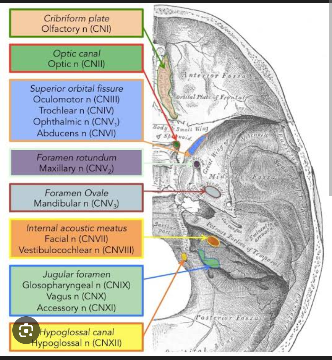
Head Indian Medical PG Question 2: Which of the following structures is located within the cavernous sinus?
- A. Maxillary division of V nerve
- B. Mandibular division of V nerve
- C. Internal carotid artery (Correct Answer)
- D. Facial nerve
Head Explanation: ***Internal carotid artery***
- The **internal carotid artery** passes **through the lumen** of the cavernous sinus, which is a dural venous sinus located on either side of the sella turcica.
- Along with the **abducens nerve (CN VI)**, the internal carotid artery is one of only two structures that passes directly through the cavernous sinus cavity itself.
- This is the **most accurate answer** as it traverses the actual sinus space, not just the wall.
*Maxillary division of V nerve*
- The **maxillary division of the trigeminal nerve (V2)** runs within the **lateral wall** of the cavernous sinus, not through its lumen.
- While technically "within" the sinus structure, it is embedded in the dural wall rather than passing through the blood-filled cavity.
- This nerve exits the skull through the **foramen rotundum**.
- Other nerves in the lateral wall include **CN III, CN IV, and V1**.
*Mandibular division of V nerve*
- The **mandibular division of the trigeminal nerve (V3)** does not pass through or near the cavernous sinus.
- It exits the middle cranial fossa directly via the **foramen ovale**, positioned inferior and separate from the cavernous sinus.
- V3 is the only division of the trigeminal nerve that does not have any relationship with the cavernous sinus.
*Facial nerve*
- The **facial nerve (CN VII)** has no anatomical relationship with the cavernous sinus.
- It enters the temporal bone through the **internal acoustic meatus**, travels through the facial canal, and exits via the **stylomastoid foramen**.
- Its course is entirely separate from the cavernous sinus region.
Head Indian Medical PG Question 3: Neurological status is assessed under which step of ABCDE of trauma care?
- A. C - Circulation with haemorrhage control
- B. E - Exposure: completely undress the patient and assess for other injuries
- C. B - Breathing and ventilation
- D. D - Disability: neurological status (Correct Answer)
Head Explanation: ***D - Disability: neurological status***
- The "D" in ABCDE trauma assessment specifically stands for **Disability**, which involves a rapid assessment of the patient's **neurological status**.
- This step typically includes evaluating **level of consciousness** using tools like the AVPU scale (Alert, Voice, Pain, Unresponsive) or the Glasgow Coma Scale (GCS), assessing pupillary response, and identifying any gross motor deficits.
*C - Circulation with haemorrhage control*
- This step focuses on assessing and managing **blood flow**, including evaluating heart rate, blood pressure, capillary refill, and controlling any sources of external hemorrhage.
- While neurological issues can result from poor circulation, the primary assessment of the nervous system itself is not performed here.
*E - Exposure: completely undress the patient and assess for other injuries*
- This final step involves a thorough **inspection of the entire body** to identify hidden injuries, such as bruising, lacerations, or deformities, while simultaneously ensuring temperature regulation.
- It is for overall physical assessment, not for initial neurological evaluation.
*B - Breathing and ventilation*
- This step involves assessing the patient's **respiratory effort**, checking for symmetrical chest rise, listening to breath sounds, and intervening to ensure adequate oxygenation and ventilation.
- While critical for brain function, this step focuses on the respiratory system, not the direct assessment of neurological function.
Head Indian Medical PG Question 4: In cases of severe head trauma, at what GCS is endotracheal intubation advised?
- A. 12
- B. <=8 (Correct Answer)
- C. 10
- D. <=3
Head Explanation: ***<=8***
- A **Glasgow Coma Scale (GCS) score of 8 or less** indicates significantly impaired consciousness, putting the patient at high risk for **airway compromise** and **aspiration**.
- **Endotracheal intubation** is advised to protect the airway, ensure adequate ventilation, and facilitate neurological assessment and management in these critically ill patients.
- This is the standard **"rule of 8"** used in trauma management protocols worldwide.
*12*
- A GCS score of 12, while indicating some level of altered consciousness, is generally **not low enough** to mandate immediate endotracheal intubation solely based on GCS criteria.
- Patients with this GCS may still be able to **maintain their airway** and have a **gag reflex** intact, though close monitoring is crucial.
*10*
- A GCS score of 10 suggests moderate head injury and **altered mental status**, but generally, the patient can still **protect their airway** adequately.
- While careful monitoring is essential, intubation is usually not indicated unless there are **other signs of respiratory compromise** or impending deterioration.
*<=3*
- A GCS score of 3 is the **lowest possible score**, indicating **deep coma** and severe neurological impairment, which would certainly warrant intubation.
- However, this option is **too restrictive** as it would exclude patients with **GCS 4-8 who also require intubation** for airway protection.
- The correct threshold is **GCS ≤8**, not just the most severe cases.
Head Indian Medical PG Question 5: The infratentorial dura is supplied by branches of the ___?
- A. Accessory nerve and upper cervical nerves
- B. Only vagus nerve
- C. Upper cervical spinal nerves and vagus nerve (Correct Answer)
- D. Only upper cervical nerves
Head Explanation: ***Upper cervical spinal nerves and vagus nerve***
- The **infratentorial dura mater**, particularly the posterior fossa, receives its sensory innervation primarily from the **recurrent meningeal branches** of the upper cervical spinal nerves (C1-C3), which ascend through the foramen magnum.
- The **vagus nerve (CN X)** also contributes to the sensory supply of the infratentorial dura, specifically to the posterior fossa, through its sensory branches.
*Accessory nerve and upper cervical nerves*
- The **accessory nerve (CN XI)** is primarily a motor nerve, responsible for innervating the sternocleidomastoid and trapezius muscles, and does not directly supply the dura mater.
- While upper cervical nerves do contribute, the **vagus nerve** is also a significant contributor to infratentorial dural innervation.
*Only vagus nerve*
- While the **vagus nerve (CN X)** does contribute to the sensory innervation of the infratentorial dura, it is not the sole source.
- The **upper cervical spinal nerves** also play a crucial role in providing sensory fibers to this region.
*Only upper cervical nerves*
- The **upper cervical spinal nerves** (C1-C3) are indeed a significant source of innervation for the infratentorial dura mater.
- However, the **vagus nerve (CN X)** also provides sensory branches to this region, making the answer "only upper cervical nerves" incomplete.
More Head Indian Medical PG questions available in the OnCourse app. Practice MCQs, flashcards, and get detailed explanations.