
Systemic Pathology — MCQs

On this page
A 34-year-old Caucasian female presents with truncal obesity, a rounded "moon face", and a "buffalo hump". Serum analysis shows hyperglycemia. It is determined that a pituitary adenoma is the cause of these symptoms. Adrenal examination is expected to show?
A 3-year-old girl presents with her mother for a well-child checkup. Recent laboratory data has demonstrated a persistent normocytic anemia. Her mother denies any previous history of blood clots in her past, but she says that her mother has also had to be treated for pulmonary embolism in the recent past, and her brother has had to deal with anemia his entire life. The patient's past medical history is noncontributory other than frequent middle ear infections. The vital signs upon arrival include: temperature, 36.7°C (98.0°F); blood pressure, 106/74 mm Hg; heart rate, 111/min and regular; and respiratory rate, 17/min. On physical examination, her pulses are bounding and fingernails are pale, but breath sounds remain clear. Oxygen saturation was initially 91% on room air and electrocardiogram (ECG) shows sinus tachycardia. The patient's primary care physician orders a peripheral blood smear to further evaluate this finding, and preliminary results show a hemolytic anemia. Based on the clinical presentation and laboratory findings, which of the following pathophysiologic mechanisms is most likely responsible for this patient's condition?
A pathologist receives a skin biopsy specimen from a patient who is suspected to have developed graft-versus-host disease (GVHD) following allogeneic stem-cell transplantation. The treating physician informs the pathologist that he is specifically concerned about the diagnosis as the patient developed skin lesions on the 90th-day post-transplantation and therefore, by definition, it should be considered a case of acute GVHD. However, the lesions clinically appear like those of chronic GVHD. The pathologist examines the slide under the microscope and confirms the diagnosis of chronic GVHD. Which of the following findings on skin biopsy is most likely to have helped the pathologist to confirm the diagnosis?
A 57-year-old man with diabetes mellitus type 2 presents for a routine follow-up. His blood glucose levels have been inconsistently controlled with metformin and lifestyle modifications since his diagnosis 3 years ago. He is currently is on metformin and diet control with exercise. The vital signs are as follows a blood pressure of 122/82 mm Hg, a pulse of 83/min, a temperature of 36.3°C (97.4°F), and a respiratory rate of 10/min. At this current visit, the urinalysis results are as follows: pH 6.2 Color light yellow RBC none WBC none Protein 4+ Cast none Glucose absent Crystal none Ketone absent Nitrite absent 24-h urine protein excretion 3.7 g The urine albumin loss mapping shows: Urine albumin loss/24h current: 215 mg Urine albumin loss/24h 3 months ago: 28 mg The blood sugar analysis shows: Fasting blood sugar 153 mg/dL Post-prandial blood sugar 225 mg/dL HbA1c 7.4% Which of the following best describes the expected microscopic finding on renal biopsy?
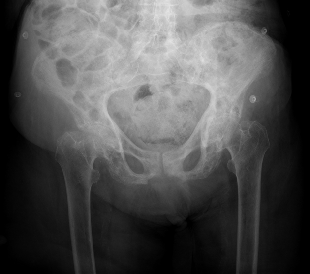
A 67-year-old man presents to the physician because of low-back pain for 6 months. The pain is more localized to the left lower back and sacral area. It is constant without any radiation to the leg. He has no significant past medical history. He takes ibuprofen for pain control. His father developed a bone disease at 60 years of age and subsequently had a fracture in the spine and another in the lower leg. The patient’s vital signs are within normal limits. The neurologic examination shows no focal findings. He has mild tenderness on deep palpation of the left pelvis. The physical examination of the lower extremities shows no abnormalities other than bowed legs. A radiograph of the pelvis is shown in the image. Which of the following serum tests is the most important initial diagnostic study?
A 68-year-old man presents with blisters on the flexor surfaces of his arms and legs. He notes that the lesions appeared 2 days ago and have not improved. He says that he has had similar blisters in the past but has not sought medical attention until now. The man has no significant past medical history. He is afebrile and his vital signs are within normal limits. On physical examination, there are tense bullae present on the flexor surfaces of his arms and legs. Biopsy of a lesion and histopathologic examination reveal a subepidermal blister with a polymorphous but predominantly eosinophilic infiltrate. Which of the following is the best next diagnostic step in this patient?
A 67-year-old man presents to his physician for a follow-up examination. During his last visit 1 month ago, splenomegaly was detected. He has had night sweats for the past several months and has lost 5 kg (11 lb) unintentionally during this period. He has no history of severe illness and takes no medications. The vital signs are within normal limits. The examination shows no abnormalities other than splenomegaly. The laboratory studies show the following: Hemoglobin 9 g/dL Mean corpuscular volume 95 μm3 Leukocyte count 12,000/mm3 Platelet count 260,000/mm3 Ultrasound shows a spleen size of 15 cm and mild hepatomegaly. A peripheral blood smear shows teardrop-shaped and nucleated red blood cells (RBCs) and immature myeloid cells. The marrow is very difficult to aspirate but reveals hyperplasia of all 3 lineages. The tartrate-resistant acid phosphatase (TRAP) test is negative. Clonal marrow plasma cells are not seen. JAK-2 is positive. The cytogenetic analysis is negative for translocation between chromosomes 9 and 22. Which of the following is the most likely diagnosis?
A 45-year-old African American man presents with nausea and severe abdominal pain. He denies vomiting. He says that, 2 days ago, his divorce was finalized, so he went to a bar and had multiple shots of tequila and vodka. This morning, upon waking, he noticed his urine was red, which lasted throughout the day. The patient denies any history of similar symptoms. Past medical history is significant for low blood counts diagnosed on routine laboratory work 6 months ago, which was not followed up due to the stress of the divorce. A review of systems is significant for erectile dysfunction and chronic fatigue. His temperature is 37.2°C (99.0°F), the heart rate is 90/min, the blood pressure is 136/88 mm Hg, and the respiratory rate is 20/min. Physical examination shows scleral icterus. Mucous membranes are pale. Cardiac auscultation reveals a systolic flow murmur loudest along the left sternal border. There is moderate right upper quadrant abdominal tenderness with no rebound or guarding. The remainder of the exam is unremarkable. Laboratory findings are significant for the following: Hematocrit 27% Mean corpuscular volume 81 µm3 Leukocytes 6,000/mm3 Platelets 130,000/µL Haptoglobin 30 mg/dL (50–150 mg/dL) Reticulocyte count 3% Total bilirubin 7.1 mg/dL LDH 766 U/L AST 150 U/L ALT 195 U/L HbA1 96% HbA2 2% HbF 2% CD55 50% of expected The peripheral smear is unremarkable. Which of the following would be the most likely cause of mortality given this patient’s likely diagnosis?
A 55-year-old African American male presents to his primary care physician with complaints of persistent back pain and fatigue over 12 months. Physical examination reveals a blood pressure of 190/150 mm Hg, and laboratory tests reveal hyperlipidemia and a serum creatinine level of 3.0 mg/dL. 4.5 g of protein are excreted in the urine over 24 hours. Renal biopsy shows eosinophilic, acellular material in the glomerular tuft and capillary walls that display apple green-colored birefringence in polarized light upon Congo red tissue staining. The patient most likely suffers from which of the following:
A 30-year-old woman was brought in by ambulance after being struck by a truck while crossing the street. She has lost a large volume of blood, and a transfusion of packed RBCs is indicated. The patient’s blood type is confirmed to be AB+. She is to be given two units of packed red blood cells (RBCs). Which of the following type(s) of packed RBCs would be safe to transfuse into this patient?
Practice by Chapter
Liver pathology (hepatitis, cirrhosis)
Practice Questions
Gallbladder and biliary tract disorders
Practice Questions
Pancreatic diseases
Practice Questions
Kidney diseases
Practice Questions
Male reproductive pathology
Practice Questions
Female reproductive pathology
Practice Questions
Breast pathology
Practice Questions
Endocrine pathology
Practice Questions
Bone and joint pathology
Practice Questions
Skeletal muscle diseases
Practice Questions
Peripheral nerve disorders
Practice Questions
Soft tissue tumors
Practice Questions
Head and neck pathology
Practice Questions
Want unlimited practice?
Get full access to all questions, explanations, and performance tracking.
Scan to download app