
Melanocytic nevi — MCQs

A 60-year-old white man with a past medical history significant for hypertension and hyperlipidemia presents to his family medicine physician with concerns about a 'spot' on his ear. He has been a construction worker for 35 years and spends most of his time outside. His family history is insignificant. On physical examination, there is a dark lesion on his left ear. The patient states that he has always had a mole in this location but that it has recently become much larger. A review of systems is otherwise negative. Which of the following lesion characteristics would be MOST reassuring among the given options?
A 38-year-old man presents to the outpatient clinic for an annual employee health checkup. He does not have any complaints at the moment except for skin changes, as seen in the following image. He denies any history of trauma. His medical history is insignificant. His family history is negative for any skin disorders or autoimmune disease. He is a non-smoker and does not drink alcohol. Which of the following is the most likely mechanism for this presentation?

A 53-year-old farmer presents to the clinic for evaluation of a pigmented lesion on his arm. He states that he first noticed the lesion last year, but he believes that it has been slowly growing in size. He otherwise does not have any complaints and is generally healthy. Which of the following findings on physical exam would suggest a malignant diagnosis?
A 28-year-old patient presents to a medical office for a consultation regarding a mole on her nose that is increasing in size. She also complains of frequent headaches, which she associates with stress on the job. She works as a civil engineer and spends much of her time outside. Her past medical history is positive for bronchial asthma; nevertheless, her vitals are stable. The mole is 8 mm in diameter, has irregular borders, and is brown in color. A biopsy is performed and sent for genetic analysis. A mutation is found. A mutation in which gene is characteristic of this patient’s main diagnosis?
A 52-year-old woman sees you in your office with a complaint of new-onset headaches over the past few weeks. On exam, you find a 2 x 2 cm dark, irregularly shaped, pigmented lesion on her back. She is concerned because her father recently passed away from skin cancer. What tissue type most directly gives rise to the lesion this patient is experiencing?
As part of a clinical research study, microscopic analysis of tissues obtained from surgical specimens is performed. Some of these tissues have microscopic findings of an increase in the size of numerous cells within the tissue with an increase in the amount of cytoplasm, but the nuclei are uniform in size. Which of the following processes shows such microscopic findings?
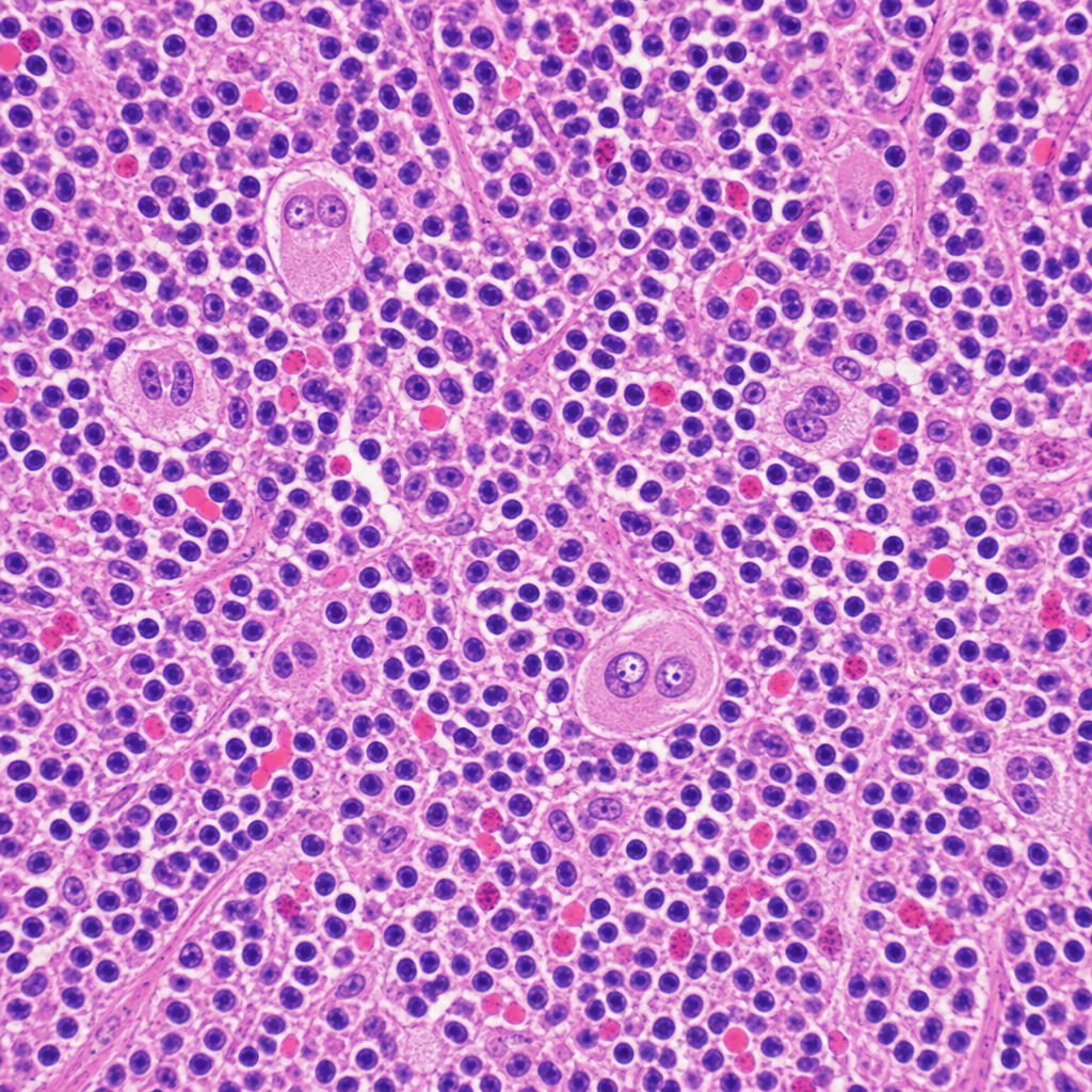
A 34-year-old woman presents with fatigue, night sweats, and a painless cervical lymph node mass for 3 months. Excisional biopsy of the lymph node is performed. The photomicrograph demonstrates a mixed cellular infiltrate with scattered large binucleated cells possessing prominent eosinophilic 'owl-eye' nucleoli, set against a background of lymphocytes, plasma cells, eosinophils, and fibroblasts. Immunohistochemical staining of the large cells is positive for CD15 and CD30 and negative for CD45. Which of the following best describes the cell of origin of the large binucleated cells seen in this lesion?
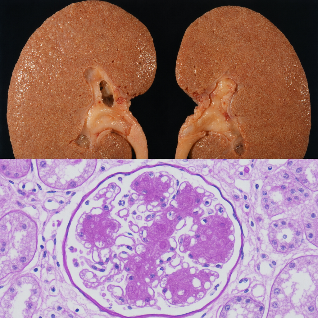
A 67-year-old man with a 20-year history of poorly controlled type 2 diabetes mellitus dies of a myocardial infarction. At autopsy, the kidneys are symmetrically enlarged with a granular cortical surface. PAS staining of the renal cortex shows PAS-positive ovoid deposits within the mesangium of glomeruli, compressing the adjacent capillary loops. Which of the following best describes the composition and pathogenetic mechanism of these deposits?
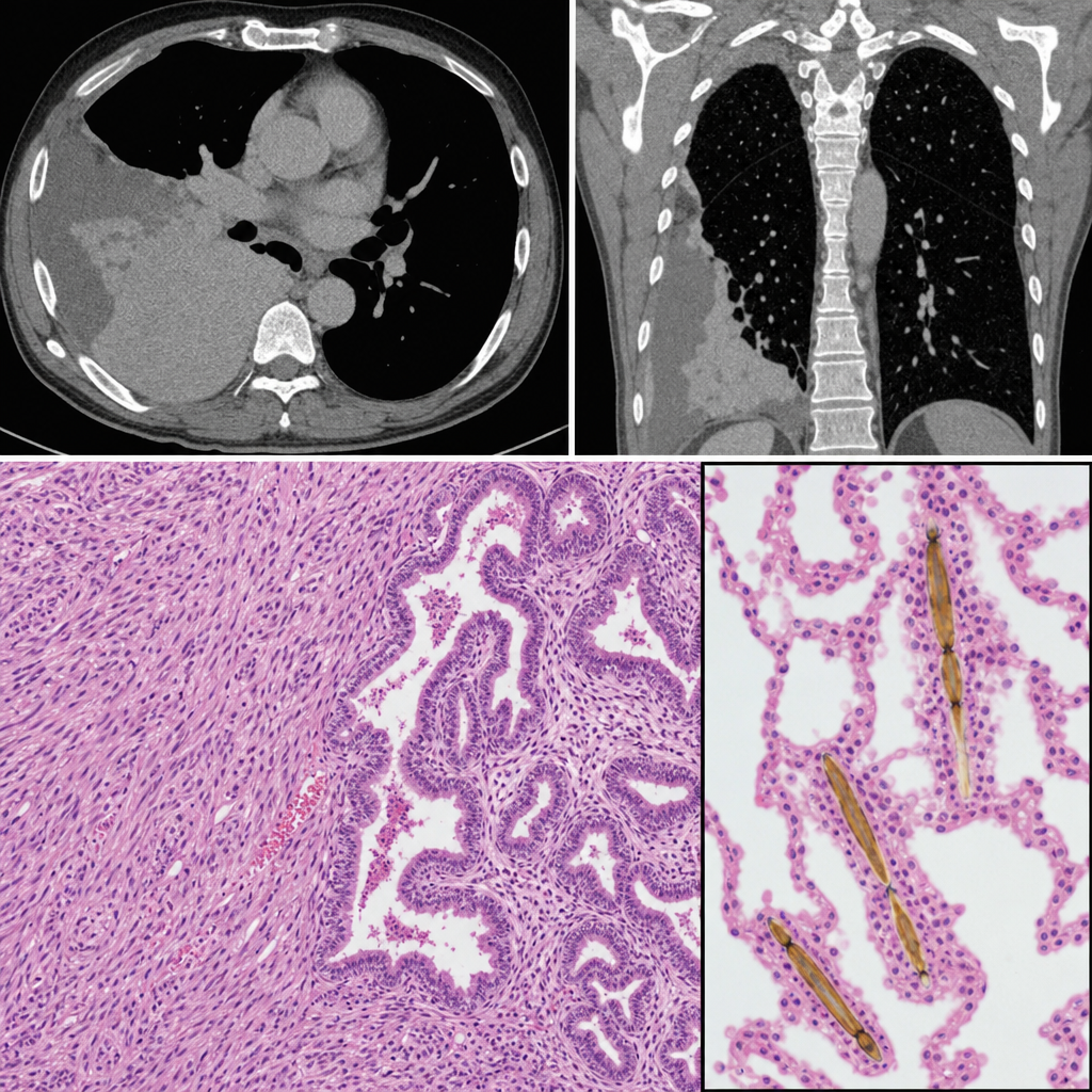
A 58-year-old man with a 40-pack-year smoking history presents with progressive dyspnea and a dry cough. Chest CT reveals bilateral pleural thickening and a right pleural mass. Thoracoscopic biopsy is performed. The photomicrograph shows a biphasic tumor with epithelioid cells forming tubulopapillary structures intermixed with a spindle-cell sarcomatoid component. Elongated, golden-brown, beaded structures with a translucent core are identified within the adjacent lung parenchyma, each coated with iron-protein complexes. Which of the following structures are most specifically represented by the coated inclusions seen in this specimen?
A 50-year-old woman with rheumatoid arthritis on methotrexate develops rapidly progressive painful ulcers on her legs with violaceous undermined borders. Biopsy shows neutrophilic dermal infiltrate with areas of necrosis, but no vasculitis or infection. Wound cultures are negative. Despite debridement, the ulcers worsen. C-ANCA and P-ANCA are negative. Evaluate the diagnosis and determine the management that addresses both the cutaneous condition and systemic disease.
Want unlimited practice?
Get full access to all questions, explanations, and performance tracking.
Scan to download app