
Dermal tumors and proliferations — MCQs

A 57-year-old post-menopausal woman comes to the physician because of intermittent, bloody post-coital vaginal discharge for the past month. She does not have pain with intercourse. Eleven years ago, she had LSIL on a routine Pap smear and testing for high-risk HPV strains was positive. Colposcopy showed CIN 1. She has not returned for follow-up Pap smears since then. She is sexually active with her husband only, and they do not use condoms. She has smoked half a pack of cigarettes per day for the past 25 years and does not drink alcohol. On speculum exam, a 1.4 cm, erythematous exophytic mass with ulceration is noted on the posterior wall of the upper third of the vagina. Which of the following is the most probable histopathology of this mass?
A 59-year-old man with chronic hepatitis C infection comes to the physician because of a 2-week history of ankle pain and nonpruritic skin lesions on his legs. He does not recall recent trauma or injury. He has not received treatment for hepatitis. Examination shows diffuse, violaceous lesions on both lower extremities. The lesions are 4–7 mm in size, slightly raised, and do not blanch with pressure. These skin lesions are best classified as which of the following?
A 6-year-old boy is brought to the physician by his parents because of right lower extremity weakness, worsening headaches, abdominal pain, dark urine, and a 5-kg (11-lb) weight loss for the past 2 months. His teachers report that he has not been paying attention in class and his grades have been worsening. He has a history of infantile seizures. Physical examination shows a palpable abdominal mass and left costovertebral angle tenderness. Neurological exam shows decreased strength of the right lower limb. He has several acne-like angiofibromas around the nose and cheeks. Further evaluation is most likely to show which of the following?
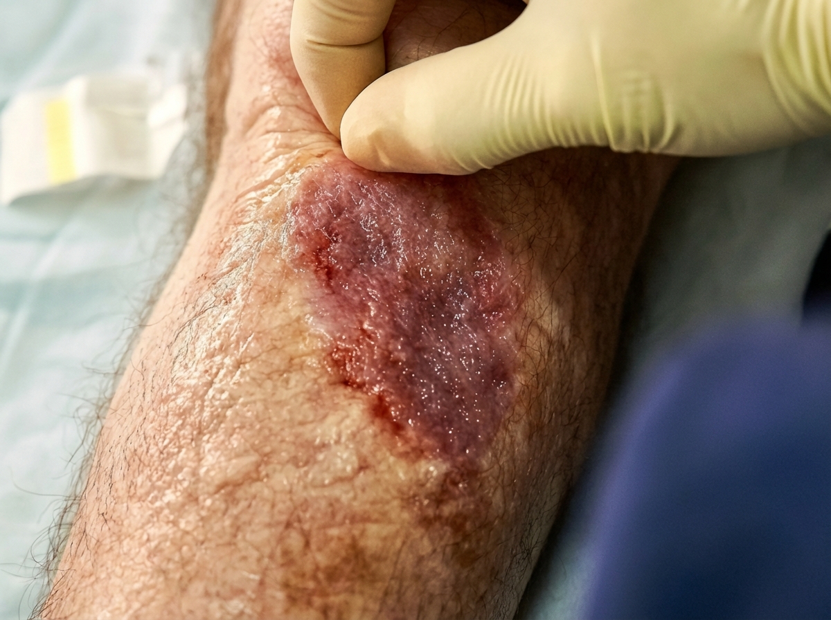
A 57-year-old man presents with a large wound on his right lower leg that has been present for 6 months as shown in the picture. He has had chronically swollen legs for over 10 years. His mother and brother had similar problems with their legs. He had a documented deep vein thrombosis (DVT) in the affected leg 5 years earlier, but has no other past medical history. He has a blood pressure of 126/84 and heart rate of 62/min. Which of the following is the most likely diagnosis?
A 23-year-old man comes to the physician for evaluation of decreased hearing, dizziness, and ringing in his right ear for the past 6 months. Physical examination shows multiple soft, yellow plaques and papules on his arms, chest, and back. There is sensorineural hearing loss and weakness of facial muscles bilaterally. His gait is unsteady. An MRI of the brain shows a 3-cm mass near the right internal auditory meatus and a 2-cm mass at the left cerebellopontine angle. The abnormal cells in these masses are most likely derived from which of the following embryological structures?
An investigator studying the molecular characteristics of various malignant cell lines collects tissue samples from several families with a known mutation in the TP53 tumor suppressor gene. Immunohistochemical testing performed on one of the cell samples stains positive for desmin. This sample was most likely obtained from which of the following neoplasms?
A 48-year-old man comes to the physician because of a skin lesion on his nose and in his mouth. The lesions have been gradually increasing in size and are not painful or pruritic. Two months ago, he was treated for esophageal candidiasis. Physical examination shows one pinkish-brown papule on the right wing of the nose and two similar nodular lesions on the hard palate and buccal mucosa. A biopsy of one of the lesions shows spindle-shaped endothelial cells and infiltration of lymphocytes, plasma cells, and macrophages. Which of the following is the most likely causal organism of this patient's condition?
A 45-year-old man comes to the emergency department because of chills and numerous skin lesions for 1 week. He has also had watery diarrhea, nausea, and abdominal pain for the past 2 weeks. The skin lesions are nonpruritic and painless. He was diagnosed with HIV infection approximately 20 years ago. He has not taken any medications for over 5 years. He sleeps in homeless shelters and parks. Vital signs are within normal limits. Examination shows several bright red, friable nodules on his face, trunk, extremities. The liver is palpated 3 cm below the right costal margin. His CD4+ T-lymphocyte count is 180/mm3 (N ≥ 500). A rapid plasma reagin test is negative. Abdominal ultrasonography shows hepatomegaly and a single intrahepatic 1.0 x 1.2-cm hypodense lesion. Biopsy of a skin lesion shows vascular proliferation and abundant neutrophils. Which of the following is the most likely causal organism?
A 52-year-old man presents to the office for evaluation of a 'weird rash' that appeared over his torso last week. The patient states that the rash just seemed to appear, but denies itching, pain, or exposure. On physical examination, the patient has multiple light brown-colored flat plaques on the torso. They appear to be 'stuck on' but do not have associated erythema or swelling. What condition is this clinical presentation most likely associated with?
Two weeks after undergoing low anterior resection for rectal cancer, a 52-year-old man comes to the physician because of swelling in both feet. He has not had any fever, chills, or shortness of breath. His temperature is 36°C (96.8°F) and pulse is 88/min. Physical examination shows a normal thyroid and no jugular venous distention. Examination of the lower extremities shows bilateral non-pitting edema that extends from the feet to the lower thigh, with deep flexion creases. His skin is warm and dry, and there is no erythema or rash. Microscopic examination of the interstitial space in this patient's lower extremities would be most likely to show the presence of which of the following?
Want unlimited practice?
Get full access to all questions, explanations, and performance tracking.
Scan to download app