
Pelvic radiologic landmarks — MCQs

A 36-year-old male is taken to the emergency room after jumping from a building. Bilateral fractures to the femur were stabilized at the scene by emergency medical technicians. The patient is lucid upon questioning and his vitals are stable. Pain only at his hips was elicited. Cervical exam was not performed. What is the best imaging study for this patient?
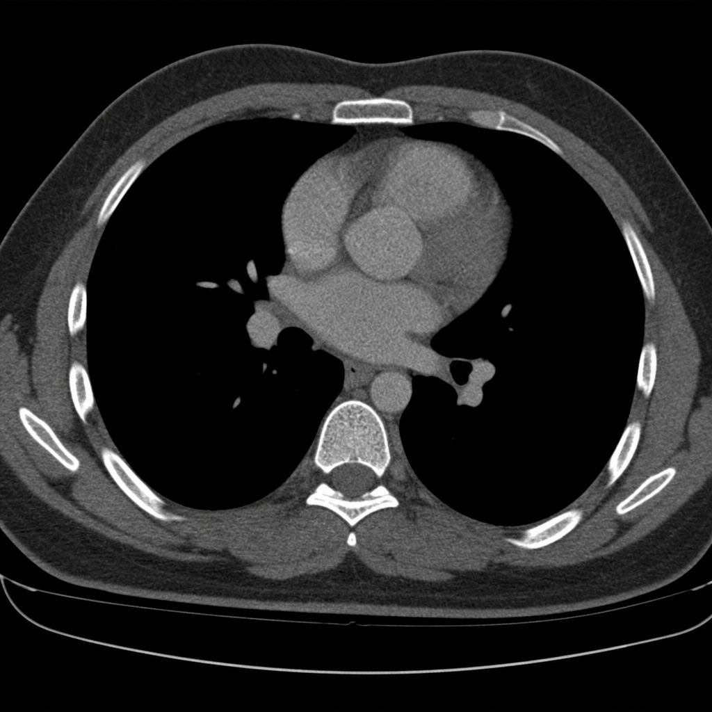
A 40-year-old woman is brought to the emergency department by a paramedic team from the scene of a motor vehicle accident where she was the driver. The patient was restrained by a seat belt and was unconscious at the scene. On physical examination, the patient appears to have multiple injuries involving the trunk and extremities. There are no penetrating injuries to the chest. As part of her trauma workup, a CT scan of the chest is ordered. At what vertebral level of the thorax is this image from?
An 80-year-old woman is brought to the emergency department for left hip pain 30 minutes after she fell while walking around in her room. Examination shows left groin tenderness. The range of motion of the left hip is limited because of pain. An x-ray of the hip shows a linear fracture of the left femoral neck with slight posterior displacement of the femur. Which of the following arteries was most likely damaged in the patient's fall?
A 69-year-old woman presents with pain in her hip and groin. She states that the pain is present in the morning, and by the end of the day it is nearly unbearable. Her past medical history is notable for a treated episode of acute renal failure, diabetes mellitus, obesity, and hypertension. Her current medications include losartan, metformin, insulin, and ibuprofen. The patient recently started taking high doses of vitamin D as she believes that it could help her symptoms. She also states that she recently fell off the treadmill while exercising at the gym. On physical exam you note an obese woman. There is pain, decreased range of motion, and crepitus on physical exam of her right hip. The patient points to the areas that cause her pain stating that it is mostly over the groin. The patient's skin turgor reveals tenting. Radiography is ordered. Which of the following is most likely to be found on radiography?
A 40-year-old woman comes to the physician because of a 2-week history of anal pain that occurs during defecation and lasts for several hours. She reports that she often strains during defecation and sees bright red blood on toilet paper after wiping. She typically has 3 bowel movements per week. Physical examination shows a longitudinal, perianal tear. This patient's symptoms are most likely caused by tissue injury in which of the following locations?
An MRI of a patient with low back pain reveals compression of the L5 nerve root. Which of the following muscles would most likely show weakness during physical examination?
A 45-year-old man is brought to the emergency department following a motor vehicle collision. He reports right hip pain and numbness along the right thigh. Physical examination shows decreased sensation to light touch over a small area of the proximal medial thigh. X-rays of the pelvis show a displaced pelvic ring fracture. Further evaluation of this patient is most likely to show which of the following findings?
A 35-year-old man comes to the physician because of a 3-month history of intermittent right lateral hip pain that radiates to the thigh. Climbing stairs and lying on his right side aggravates the pain. Examination shows tenderness to palpation over the upper lateral part of the right thigh. There is no swelling. When the patient is asked to abduct the right leg against resistance, tenderness is noted. An x-ray of the pelvis shows no abnormalities. Which of the following structures is the most likely source of this patient's pain?
A 25-year-old man presents with progressive weakness and urinary retention. MRI of the spine shows an intramedullary lesion from T10-T12 with expansion of the spinal cord and syrinx formation. The conus medullaris is identified at the L1-L2 level (normal: L1-L2). The filum terminale appears thickened at 3 mm. CSF flow study shows obstruction at the lesion site. Evaluate these radiologic landmarks and their relationships to determine the neurological level most likely affected.
A 58-year-old woman with breast cancer undergoes staging CT. The scan shows a solitary 2 cm lesion in the liver at the junction of segments IVa, V, and VIII, directly adjacent to the middle hepatic vein. PET scan shows FDG avidity. The oncologist requests evaluation for surgical resection. The radiologist notes the lesion's relationship to the portal vein bifurcation (Cantlie's line). Evaluate the radiologic anatomical landmarks to determine resectability and surgical approach.
Want unlimited practice?
Get full access to all questions, explanations, and performance tracking.
Scan to download app