
Radiographic Anatomy of Extremities — MCQs

What condition is primarily diagnosed using Von Rosen's view?
In a case of recurrent anterior dislocation of the shoulder, posterolateral lesions were found on radiological examination. What are these lesions?
The commonly injured carpal bone next to the scaphoid is:
A 40-year-old presents with chronic shoulder pain and restricted ROM. X-ray shows decreased joint space. Most likely diagnosis?
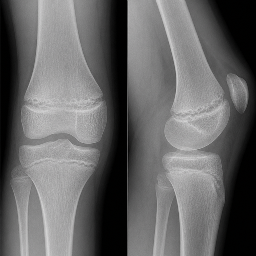
The image shows a pediatric fracture involving the growth plate. Which classification system and stage best describes this fracture?
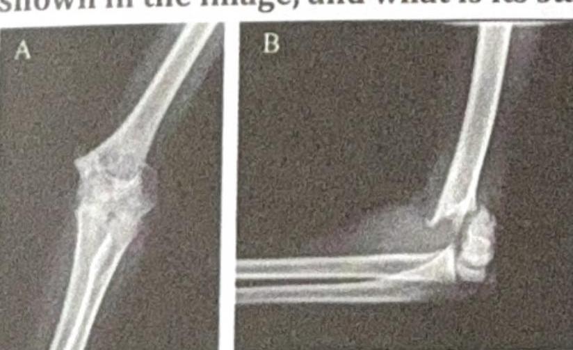
Which Salter-Harris fracture type involves a metaphyseal fragment?
A radiograph is obtained from a child with scoliosis. What is the name of the angle used to measure spinal curvature?
A 14-year-old presents with knee pain and limping. X-ray shows widened irregular growth plate at distal femur. What is the diagnosis?
A patient came with history of fall and on examination there was tenderness between the extensor pollicis longus and brevis. The likely lesion is
The best view to visualize zygomatic arches is
Want unlimited practice?
Get full access to all questions, explanations, and performance tracking.
Scan to download app