
Radiographic Anatomy of Chest — MCQs

This 23-year-old man was involved in a motor vehicle accident. He presents with shortness of breath and chest pain. On examination, there is decreased breath sound on the right side and subcutaneous emphysema. Chest X-ray shows a deep, lucent right costophrenic angle. What is the diagnosis?
"Hour-glass" shape of the chest and "tri-radiate pelvis" are seen radiologically in -
Lower limit of the left crus of the diaphragm is at which vertebral level?
While performing drainage of fluid from the pleural cavity, the needle is introduced through all of the following structures except which one?
PA view of chest X-ray is given here. What is the diagnosis?

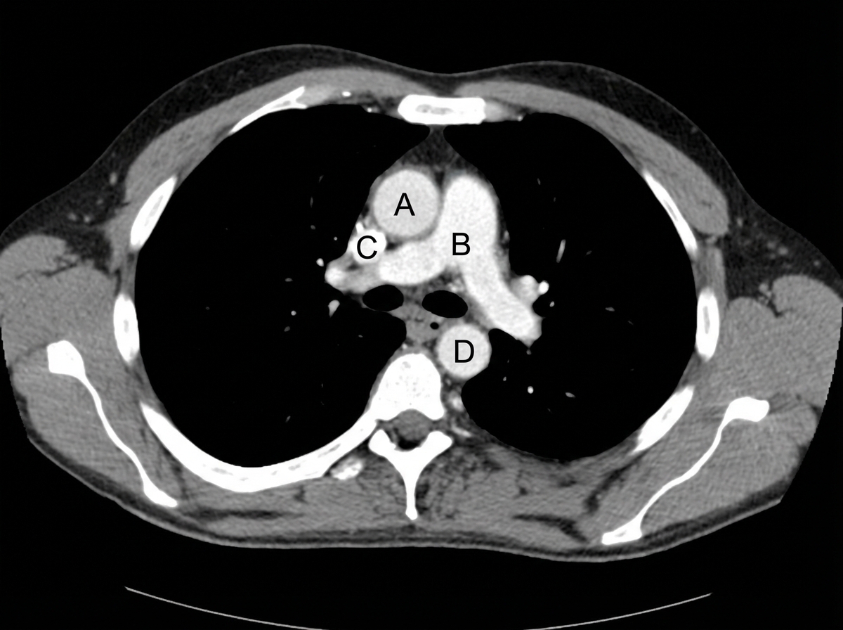
Identify the labeled structures correctly in the axial CT image of the thorax
Which airway structure enters the lung at the hilum?
A 10yr old boy with a known case of nephrotic syndrome since 4 years on treatment brought to the pediatric OPD with chief complaint of difficulty in breathing. There is no history of fever. On examination, respiratory system was normal except slightly reduced breath sounds on right infra-axillary region. Paediatrician thinks of pleural effusion. What is next best modality of investigation to detect pleural effusion?
Match the following: A) Caplan syndrome- 1) Found first in coal worker B) Asbestosis- 2) Upper lobe predominance C) Mesothelioma- 3) Involves lower lobe D) Sarcoidosis- 4) Pleural effusion is seen
Carina is situated at which level?
Want unlimited practice?
Get full access to all questions, explanations, and performance tracking.
Scan to download app