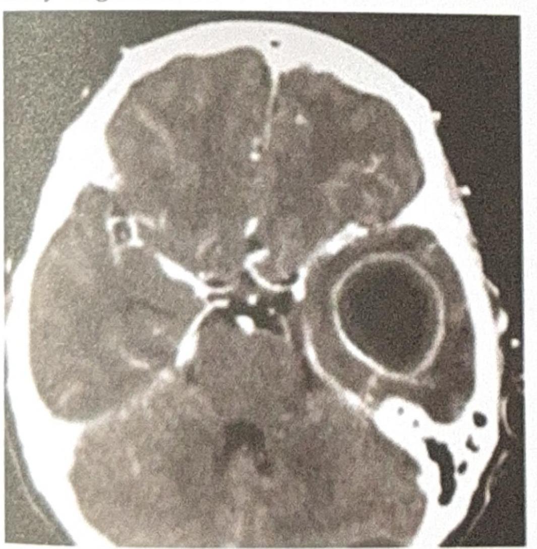
A patient presents with ear discharge. The CT image is shown below. Based on the clinical presentation and imaging, what is the most likely diagnosis?

An 34-year-old male HIV patient on c-A presents with seizures and a unilateral facial nerve palsy. The CT scan shows a ring-enhancing lesion. What is the best treatment?

Symptomatic neonatal CNS involvement is most commonly seen in which group of congenital intrauterine infections?
All of the following statements are true regarding central nervous system infections, except:
Which of the following statements is true regarding HSV encephalitis?
A leukemia patient who has undergone multiple courses of chemotherapy develops herpes simplex encephalitis. Which of the following would you expect a CT scan of the patient's brain to show?
A 65-year-old diabetic man presents with black necrotic tissue on his palate. What is the most likely causative organism?
Which of the following is the MOST COMMON fungal cause of Mucormycosis?
A 40-year-old male presents with a history of headaches, fever, and new-onset seizures. An MRI of the brain is performed, revealing a ring-enhancing lesion with central restricted diffusion on diffusion-weighted imaging (DWI). What is the most likely diagnosis?
Which of the following conditions is most commonly associated with the "doughnut" sign seen on a brain scan?
Explanation: ***Temporal lobe abscess*** - The CT scan shows a **ring-enhancing lesion** in the **temporal lobe**, which is characteristic of a brain abscess. - **Ear discharge** (otorrhea), particularly from otitis media, is a common predisposing factor for temporal lobe abscesses due to the proximity of the middle ear and mastoid to the temporal lobe. - Otogenic brain abscesses account for a significant proportion of intracranial complications from ear infections, with the temporal lobe being the most common location. *Extradural abscess* - An **extradural abscess** would typically be located between the dura mater and the skull, often presenting as a **lenticular or biconvex collection** displacing the dura and brain, not within the brain parenchyma as seen here. - While ear infections can lead to extradural abscesses, the imaging clearly shows an intraparenchymal lesion. *Cerebellar abscess* - A **cerebellar abscess** would be located in the cerebellum (posterior fossa), which is a different anatomical location from the lesion seen in the image (which is in the supratentorial compartment, consistent with the temporal lobe). - Although ear infections can also lead to cerebellar abscesses, the lesion's position on the CT scan does not correspond to the cerebellum. *Meningitis* - **Meningitis** is an inflammation of the meninges and typically manifests on CT as **leptomeningeal enhancement**, particularly in the sulci and basal cisterns, rather than a discrete, encapsulated mass lesion like an abscess. - While ear discharge can be associated with meningitis, the imaging findings strongly point to an abscess, not diffuse meningeal inflammation.
Explanation: ***Sulphadiazine, pyrimethamine and Leucovorin*** - This combination is the standard **first-line treatment for cerebral toxoplasmosis**, which is strongly suggested by the clinical presentation (HIV patient, seizures, facial nerve palsy) and the imaging findings of **multiple ring-enhancing lesions**. - **Leucovorin** is added to prevent bone marrow suppression caused by pyrimethamine. *Albendazole with dexamethasone* - **Albendazole** is primarily used for **neurocysticercosis**, which typically presents with cystic lesions, not necessarily ring-enhancing, and the patient's HIV status makes toxoplasmosis more likely. - While **dexamethasone** may be used to reduce brain edema, it's adjunctive and not the primary antimicrobial treatment for toxoplasmosis. *Amphotericin B* - **Amphotericin B** is the mainstay treatment for **cryptococcal meningitis** and other severe fungal infections, which usually present with symptoms of meningitis and different imaging findings (e.g., hydrocephalus, gelatinous pseudocysts). - It is not effective against **Toxoplasma gondii**. *ATT with steroids* - **ATT (Anti-Tubercular Therapy)** with steroids is the treatment for **CNS tuberculosis**, which can present with ring-enhancing lesions. - However, the typical presentation for CNS tuberculosis in HIV patients often includes basilar meningitis, multiple tuberculomas, or abscesses, and toxoplasmosis is a far more common cause of ring-enhancing lesions in HIV patients with CD4 counts < 100 cells/µL.
Explanation: ***CMV and toxoplasmosis*** - Both **cytomegalovirus (CMV)** and **Toxoplasma gondii** are well-known causes of congenital infections that frequently lead to significant and symptomatic central nervous system (CNS) involvement in neonates. - Congenital CMV can cause **microcephaly**, **periventricular calcifications**, **hearing loss**, and developmental delay, while congenital toxoplasmosis can result in **hydrocephalus**, **intracranial calcifications**, **chorioretinitis**, and seizures. *Rubella and toxoplasmosis* - While **toxoplasmosis** causes significant CNS involvement, **congenital rubella syndrome** typically presents with cataracts, heart defects (e.g., patent ductus arteriosus), and hearing loss, with CNS involvement being less consistently severe or frequently symptomatic in the immediate neonatal period compared to CMV or toxoplasmosis. - Although rubella can cause **encephalitis** or **meningoencephalitis**, these are not as common or consistently severe as the direct destructive CNS lesions seen with CMV or toxoplasmosis. *CMV and syphilis* - **CMV** is a major cause of neonatal CNS symptoms. However, **congenital syphilis** primarily affects bones, skin, and mucous membranes (e.g., "snuffles"), with CNS involvement typically presenting as **meningitis**, **hydrocephalus**, or neurodevelopmental delays, but often not as overtly symptomatic in the immediate neonatal period as CMV or toxoplasmosis. - While syphilis can cause neurosyphilis, the spectrum and severity of immediate symptomatic CNS involvement are distinct from the widespread calcifications and structural abnormalities seen with CMV or toxoplasmosis. *Rubella and HSV* - **Rubella** primarily causes classic congenital defects in eyes, ears, and heart, with CNS effects being less common and severe. - **Congenital herpes simplex virus (HSV)** infection, while causing severe CNS disease (e.g., encephalitis) when disseminated, is relatively rare overall compared to CMV and toxoplasmosis, and often presents with skin, eye, and mouth lesions first.
Explanation: ***Cytomegalovirus is a common cause of bilateral temporal lobe hemorrhagic infarction.*** - **Cytomegalovirus (CMV)** typically causes **ventriculoencephalitis or periventricular necrosis** and microglial nodules in immunocompromised patients, not bilateral temporal lobe hemorrhagic infarction. - **Herpes simplex virus type 1 (HSV-1)** is the classic infectious cause of **bilateral temporal lobe hemorrhagic infarction (necrotizing encephalitis)**. *Prions infection causes spongiform encephalopathy* - **Prions** are misfolded proteins that cause transmissible spongiform encephalopathies (TSEs), such as Creutzfeldt-Jakob disease, characterized by **neuronal loss** and vacuolation (spongiform changes). - These diseases are invariably fatal and lead to rapid neurological deterioration. *JC virus is causative agent for progressive multifocal leucoencephalopathy* - The **JC virus** specifically targets and destroys **oligodendrocytes**, the myelin-producing cells of the central nervous system. - This leads to **demyelination** in multiple areas of the brain, causing the characteristic lesions seen in progressive multifocal leukoencephalopathy (PML). *Measles virus is the causative agent for subacute sclerosing pan encephalitis (SSPE).* - **Subacute sclerosing panencephalitis (SSPE)** is a rare, fatal, progressive neurodegenerative disease caused by a persistent and defective **measles virus infection** in the brain. - It occurs years after the initial measles infection, leading to cognitive decline, seizures, and motor dysfunction.
Explanation: ***Hemorrhagic lesions are seen*** - **Hemorrhagic necrosis** of the **temporal lobes** is the pathognomonic feature of HSV encephalitis [3] - The hemorrhagic nature helps differentiate it from other viral encephalitides - Classically affects the **medial temporal lobes** and **inferior frontal lobes** bilaterally [2] - MRI shows hemorrhagic changes with mass effect in these regions *Caused by HSV-1* - While **HSV-1** causes >90% of HSV encephalitis cases in adults and children, this statement is technically correct - However, **hemorrhagic lesions** are the more distinctive pathological feature being tested - HSV-1 is the causative agent, but the hemorrhagic pathology is the key diagnostic finding *Eosinophilic inclusion bodies are seen* - **Cowdry type A intranuclear inclusion bodies** are indeed seen in HSV encephalitis [1] - These are **eosinophilic** with a clear halo around them - While true, this is a microscopic finding, whereas hemorrhagic lesions are the macroscopic hallmark *Caused by Varicella zoster virus* - VZV causes **varicella** (chickenpox) and **herpes zoster** (shingles) [2] - VZV can cause encephalitis but with different clinical and pathological features - HSV encephalitis is specifically caused by **herpes simplex virus**, not varicella zoster virus **References:** [1] Kumar V, Abbas AK, et al.. Robbins and Cotran Pathologic Basis of Disease. 9th ed. Infectious Diseases, pp. 365-366. [2] Kumar V, Abbas AK, et al.. Robbins and Cotran Pathologic Basis of Disease. 9th ed. The Central Nervous System, pp. 1278-1279. [3] Kumar V, Abbas AK, et al.. Robbins and Cotran Pathologic Basis of Disease. 9th ed. The Central Nervous System, p. 1278.
Explanation: ***Focal edema and hemorrhagic changes selectively in the temporal and frontal lobes*** - **Herpes simplex encephalitis (HSE)** characteristically causes inflammation and damage in the **temporal and frontal lobes** due to the virus's preferential tropism for these regions. - CT scans in HSE often reveal signs of **edema**, **hemorrhage**, and sometimes **mass effect**, particularly in these specific areas. *Generalized edema and hemorrhagic changes* - While encephalitis can cause edema, **generalized edema** and widespread hemorrhagic changes are less typical for HSE, which tends to have a more **localized** presentation. - This pattern might be seen in other severe forms of encephalitis or diffuse brain injury, but it's not the hallmark of **HSE**. *Focal edema and hemorrhagic changes selectively in the brainstem* - Involvement of the **brainstem** is uncommon in typical HSE; instead, the temporal and frontal lobes are the primary targets for viral replication and inflammatory response. - Brainstem encephalitis can occur with other pathogens or autoimmune conditions, but it's not the characteristic presentation of **herpes simplex encephalitis**. *Focal edema and hemorrhagic changes selectively in the basal ganglia* - Lesions predominantly in the **basal ganglia** are not characteristic of HSE, which typically spares these deep brain structures. - Basal ganglia involvement can be seen in conditions like carbon monoxide poisoning, metabolic encephalopathies, or certain deep-seated infections, but not typically in **HSE**.
Explanation: ***Mucor species*** - The presence of **black necrotic tissue** on the palate in a diabetic patient is highly suggestive of **mucormycosis**, an aggressive fungal infection caused by *Mucor* species. - **Diabetes mellitus**, particularly with ketoacidosis, is a major risk factor for mucormycosis due to impaired phagocytic function and increased iron availability. *Cryptococcus neoformans* - This fungus is primarily associated with **cryptococcal meningitis** or pneumonia, especially in immunocompromised individuals. - It does not typically cause **black necrotic lesions** on the palate. *Candida albicans* - While *Candida albicans* can cause oral infections (**thrush**), these typically present as white, creamy patches that can be scraped off, not black necrotic tissue. - Oral candidiasis is common in diabetics but does not usually involve tissue necrosis. *Aspergillus fumigatus* - *Aspergillus* species can cause invasive infections, particularly in immunocompromised patients, often affecting the lungs or sinuses. - While it can cause **necrotic lesions**, the characteristic rapid progression and specific presentation in the palate of a diabetic with black necrotic tissue points more strongly towards *Mucor*.
Explanation: ***Rhizopus oryzae*** - This is the **most common genus and species** responsible for mucormycosis infections in humans. - It belongs to the order **Mucorales**, which are characterized by broad, non-septate hyphae with a tendency to invade blood vessels. *Candida albicans* - This fungus is a common cause of **candidiasis**, which can manifest as thrush, vaginitis, or invasive candidemia. - It does not cause mucormycosis; its cellular morphology and disease presentation are distinct. *Aspergillus fumigatus* - This is the primary causative agent of **aspergillosis**, which can range from allergic reactions to invasive forms like chronic pulmonary aspergillosis or invasive aspergillosis. - While it can cause severe fungal infections, it is morphologically distinct (septate hyphae) and does not cause mucormycosis. *Cryptococcus neoformans* - This encapsulated yeast is best known for causing **cryptococcosis**, particularly **meningoencephalitis** in immunocompromised individuals. - Its disease presentation and microscopic features (yeast with budding, prominent capsule) are entirely different from those of Mucorales.
Explanation: ***Brain abscess*** - A **ring-enhancing lesion** with **central restricted diffusion** on DWI is highly characteristic of a brain abscess, due to the presence of pus containing densely packed inflammatory cells and bacteria with high viscosity. - The clinical presentation of **headaches, fever**, and **new-onset seizures** is consistent with an infectious process and increased intracranial pressure. - This combination of imaging and clinical features is pathognomonic for pyogenic brain abscess. *Glioblastoma multiforme* - While GBM can present with **ring-enhancing lesions** and seizures, it typically exhibits **facilitated diffusion** (high ADC values) on DWI due to necrotic tumor core, not restricted diffusion. - GBM is a highly infiltrative tumor with extensive **vasogenic edema**. - Fever is uncommon in GBM unless there is secondary infection. *Metastatic brain tumor* - Metastatic lesions can be **ring-enhancing** and cause seizures, but **restricted diffusion** is not typical unless there is acute hemorrhage or superimposed infection. - The presence of **fever** points away from uncomplicated metastasis. - Multiple lesions at the gray-white matter junction are more typical of metastases. *Toxoplasmosis* - Toxoplasmosis in **immunocompromised individuals** (HIV/AIDS with CD4 <100) causes **multiple ring-enhancing lesions** with predilection for basal ganglia. - Restricted diffusion is **not consistently seen** with toxoplasmosis, unlike pyogenic abscesses. - The specific DWI finding of central restricted diffusion makes brain abscess the most definitive diagnosis.
Explanation: ***Brain abscess*** - The **doughnut sign**, characterized by a **ring-enhancing lesion** with a central hypodense core, is highly suggestive of a brain abscess due to central necrosis and peripheral inflammation. - The ring enhancement often appears **thinner and smoother** in abscesses compared to tumors, and there is usually significant surrounding edema. - This sign is most **characteristically and commonly associated** with pyogenic brain abscesses. *Brain metastases* - While brain metastases can also present as **ring-enhancing lesions**, they tend to have a **thicker and more irregular wall** compared to a brain abscess. - They often present with **multiple lesions** and a known primary malignancy, which differentiates them from a solitary abscess. *Glioblastoma multiforme* - **Glioblastoma multiforme (GBM)** typically shows a **thick, irregular, and often nodular ring enhancement** due to areas of necrosis, hypervascularity, and active tumor growth. - Its enhancing rim is generally **thicker and more variable** than that of a typical abscess, and it often invades surrounding brain parenchyma. *Toxoplasmosis* - **Cerebral toxoplasmosis** can present with ring-enhancing lesions, particularly in immunocompromised patients (HIV/AIDS). - Typically shows **multiple lesions** with predilection for basal ganglia and corticomedullary junction. - The "**eccentric target sign**" (eccentric dot within the ring) is more characteristic of toxoplasmosis than the smooth doughnut sign of pyogenic abscess.
Get full access to all questions, explanations, and performance tracking.
Start For Free