Techniques used for protein expression proteomics study include:
Best imaging modality for acoustic neuroma screening
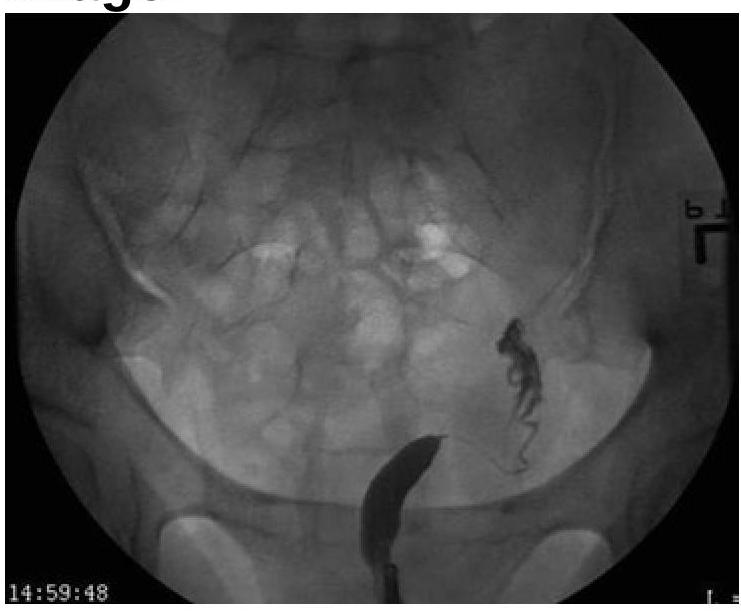
Based on the provided image, which of the following is the correct diagnosis?

Which of the following is the platinum-based chemotherapeutic agent used as first-line treatment for ovarian carcinoma?
Gold standard investigation for breast carcinoma screening in a patient with silicone breast implants
TRUS in carcinoma prostate is most useful for?
What is the full form of DICOM?
Radiation-induced necrosis can be diagnosed by:
Isotope used in bone scans:
Parameningeal Rhabdomyosarcoma is best diagnosed by:
Explanation: ***All of the options*** - All listed techniques—**Polyacrylamide Gel Electrophoresis (PAGE)**, **Gene Expression Analysis**, and **Mass Spectrometry**—are used in protein expression proteomics studies, either directly or indirectly, to analyze and quantify proteins. - The integration of these various techniques provides a comprehensive approach to understanding protein expression profiles. *PolyAcrylamide Gel Electrophoresis (PAGE)* - **PAGE** (including 1D and 2D-PAGE) is a fundamental technique for separating proteins based on their **molecular weight** and **isoelectric point**, which is crucial for visualizing and quantifying expressed proteins. - It often serves as an initial separation step before more detailed analysis, such as **mass spectrometry**. *Gene Expression Analysis (indirectly related to proteomics)* - Although **gene expression analysis** (e.g., using **RT-PCR** or **microarrays**) measures mRNA levels, it is indirectly related to proteomics because mRNA levels often **correlate with protein levels**. - It provides insights into the **transcriptional regulation** that influences protein expression, complementing direct protein analysis. *Mass Spectrometry* - **Mass spectrometry** is a powerful and widely used technique in proteomics for **identifying, quantifying, and characterizing proteins** and peptides by measuring their **mass-to-charge ratio**. - It can be used for both **discovery proteomics** (identifying novel proteins) and **targeted proteomics** (quantifying specific proteins).
Explanation: ***MRI with gadolinium*** - **Magnetic Resonance Imaging (MRI) with gadolinium contrast** is the gold standard for acoustic neuroma (vestibular schwannoma) detection due to its superior soft tissue resolution. - It effectively visualizes **small tumors** arising from the vestibular nerve within the internal auditory canal and cerebellopontine angle. *Nuclear scan* - **Nuclear scans** are generally used for assessing metabolic activity or specific tissue uptake, such as in oncology for metastasis detection or thyroid conditions. - They lack the **anatomical detail and resolution** needed to visualize small intracranial tumors like acoustic neuromas. *CT temporal bone* - **CT scans of the temporal bone** are excellent for evaluating bony structures, such as fractures or erosion of the internal auditory canal. - However, they have **limited sensitivity for soft tissue masses** and can miss small acoustic neuromas. *Plain skull X-ray* - **Plain skull X-rays** provide very limited information about soft tissues and are not useful for screening or diagnosing acoustic neuromas. - They mainly visualize **gross bony abnormalities** and cannot detect subtle pathologies within the internal auditory canal or cerebellopontine angle.
Explanation: ***Unicornuate Uterus*** - The image distinctly shows **only one fallopian tube and one rudimentary uterine horn** on the right side, indicating a unicornuate uterus. - This malformation results from the **incomplete development of one Müllerian duct**, leading to a single, banana-shaped uterine cavity. *Uterus didelphys* - This condition involves **two completely separate uteri**, each with its own cervix and vagina. - The image does not show evidence of two distinct uterine bodies or cervices. *Bicornuate Uterus* - A bicornuate uterus is characterized by **two uterine horns that fuse caudally**, creating a heart-shaped appearance with a shared cervix. - The image clearly lacks the characteristic heart shape and shows only one functional horn. *Septate uterus* - A septate uterus has a **fibrous or muscular septum** dividing the uterine cavity, while the external uterine contour remains normal. - The image does not show a septum or a normal external uterine contour with an internal division; instead, it presents with a single underdeveloped horn.
Explanation: ***Cisplatin*** - **Cisplatin** is a platinum-based chemotherapy drug that forms **DNA cross-links**, inhibiting DNA synthesis and leading to the death of rapidly dividing cells, making it highly effective against **ovarian carcinoma**. - It is a cornerstone of chemotherapy regimens for ovarian cancer, often used in combination with other agents such as paclitaxel. *Methotrexate* - **Methotrexate** is an **antimetabolite** that inhibits dihydrofolate reductase, thereby interfering with DNA synthesis. - While it is used in various cancers like leukemia, lymphoma, and some solid tumors (e.g., breast cancer, gestational trophoblastic disease), it is **not a primary recommended drug for ovarian carcinoma**. *Cyclophosphamide* - **Cyclophosphamide** is an **alkylating agent** that causes DNA damage, leading to cell death. - It is used in many cancers, including lymphoma, breast cancer, and some leukemias, but it is **not a first-line or primary agent for ovarian carcinoma** in contemporary treatment guidelines. *Dacarbazine* - **Dacarbazine** is an **alkylating agent** primarily used in the treatment of **malignant melanoma** and Hodgkin lymphoma. - It is **not indicated for the treatment of ovarian carcinoma**.
Explanation: ***MRI*** - **MRI** is considered the **gold standard** for breast cancer screening in patients with silicone breast implants due to its superior ability to visualize breast tissue through the implant and detect subtle lesions. - It offers **high sensitivity** in detecting both implant rupture and early malignancies, often providing better clarity than mammography in augmented breasts where implants can obscure tissue. *Mammography* - While a standard screening tool, **mammography** can be limited in patients with silicone implants because the implants can **obscure adjacent breast tissue**, making detection of small masses challenging. - Special views (e.g., **Eklund views**) can be used, but sensitivity is still reduced compared to MRI in augmented breasts. *CT scan* - **CT scans** are not routinely used for primary breast cancer screening due to their use of **ionizing radiation** and lower sensitivity for detecting early breast lesions compared to MRI. - CT is more commonly used for **staging** advanced cancers or evaluating complex masses detected by other modalities. *USG* - **Ultrasound (USG)** is a valuable complementary tool, especially for evaluating palpable lumps or clarifying findings from mammography, but it is **operator-dependent** and has a lower overall sensitivity for general screening compared to MRI. - It is particularly useful for differentiating between **cystic and solid masses** and detecting implant ruptures but is not the gold standard for comprehensive screening in augmented breasts.
Explanation: ***Assisting in targeted prostate biopsies*** - **TRUS** (Transrectal Ultrasound) provides real-time imaging, which is crucial for **guiding biopsy needles** accurately to suspicious areas within the prostate that may not be palpable. - This guidance increases the diagnostic yield of biopsies, ensuring samples are taken from potentially cancerous regions. *Evaluating nearby structures for involvement* - While TRUS can visualize the immediate surrounding structures like the **seminal vesicles**, its primary role is not for comprehensive staging of tumor extension outside the prostate, which is better achieved with MRI. - It helps in assessing direct invasion into seminal vesicles but has limitations for wider regional lymph node or distant metastasis evaluation. *Identifying suspicious areas in the prostate* - TRUS can identify **hypoechoic lesions** within the prostate, which are often associated with cancer, but these findings are not specific, and many benign conditions can mimic cancer. - The main utility is not solely in identifying these areas, but in using this identification to guide subsequent biopsies for definitive diagnosis. *Estimating the size of the prostate* - TRUS is highly effective for accurately measuring prostate volume, which is important for estimating PSA density and for surgical planning in benign prostatic hyperplasia (BPH). - However, while it can measure size, this is not its most diagnostically critical role in the context of carcinoma prostate when considering its unique capabilities.
Explanation: **Digital imaging and communications in medicine** - **DICOM** is the international standard for managing and transmitting medical images and related data, ensuring **interoperability** between different medical imaging equipment and systems. - Its purpose is to facilitate the storage, retrieval, management, and exchange of **medical images**, such as X-rays, CT scans, and MRIs, regardless of the vendor. *Direct imaging and colors in medicine* - This option incorrectly describes the purpose and scope of DICOM, which is broader than just "direct imaging" and "colors." - The standard focuses on the **digital nature** of medical images and the **communication** between devices. *Digital information and connectivity in medicine* - While DICOM deals with "digital information" and "connectivity," this option omits the crucial aspect of "imaging" in its description. - The primary focus of DICOM is on **medical images** and their communication. *Dependent interconnectivity in medicine* - This phrase does not accurately represent the function or the components of the **DICOM standard**. - DICOM enables **independent connectivity** and interoperability rather than dependent interconnectivity.
Explanation: ***Biopsy*** - A **biopsy** is the definitive diagnostic method for radiation-induced necrosis, allowing for histological examination of tissue to confirm necrosis and rule out residual or recurrent tumor. [1], [2] - It provides a direct view of cellular changes, identifying **necrosis, atypical cells**, and ruling out **malignancy**. *MRI* - While **MRI** can show structural changes indicative of necrosis (e.g., mass effect, edema), it often cannot definitively differentiate between **radiation necrosis** and **tumor recurrence.** [2] - It often shows **T1 hypointensity** and **T2 hyperintensity**, but these findings are not specific. *CT* - **CT scans** are useful for detecting gross changes like **mass effect** and **edema** but have limited sensitivity for distinguishing necrosis from tumor recurrence. - It may show **low-density lesions** but lacks the resolution and specificity for precise diagnosis. *PET* - **PET scans** measure metabolic activity and can help distinguish between **tumor recurrence** (high uptake) and **radiation necrosis** (low uptake) in some cases. - However, false positives can occur, as some inflammatory processes in necrosis can also show increased uptake, making it **less definitive** than a biopsy. **References:** [1] Kumar V, Abbas AK, et al.. Robbins and Cotran Pathologic Basis of Disease. 9th ed. The Central Nervous System, pp. 1307-1308. [2] Kumar V, Abbas AK, et al.. Robbins and Cotran Pathologic Basis of Disease. 9th ed. Neoplasia, pp. 340-341.
Explanation: **Technetium** - **Technetium-99m (Tc-99m)** is the most widely used isotope in bone scans due to its ideal physical characteristics, including a short half-life and suitable gamma energy for imaging. - It rapidly localizes to areas of **increased osteoblastic activity** and blood flow, making it effective for detecting bone pathologies like fractures, infections, and tumors. *Chromium* - **Chromium-51** is primarily used for **red blood cell labeling** to assess red cell mass, survival, and gastrointestinal bleeding. - It is not used for bone imaging because it does not accumulate in bone tissue and has different decay characteristics. *Selenium* - **Selenium-75** is used for imaging the **parathyroid glands** (e.g., in hyperparathyroidism) and in some studies of pancreatic function. - It does not have an affinity for bone tissue and is therefore unsuitable for bone scanning. *Gallium* - **Gallium-67** is primarily used in **inflammation and infection imaging** (Ga-67 scans) and some tumor imaging. - While it can accumulate in some bone pathologies (like osteomyelitis), it is not the primary or preferred isotope specifically for routine bone structural scans.
Explanation: ***MRI*** - **Magnetic Resonance Imaging (MRI)** provides excellent soft tissue contrast, which is crucial for visualizing the extent of **parameningeal rhabdomyosarcoma** and its relationship to critical structures like the **meninges**, **brainstem**, and **cranial nerves**. - It is superior for detecting **intracranial extension**, **bone erosion**, and assessing response to treatment, making it the preferred imaging modality for diagnosis and staging. *CT Scan* - **Computed Tomography (CT) scans** are good for evaluating bone involvement and calcifications but offer less detailed soft tissue resolution compared to MRI. - While it can identify large masses, it may miss subtle extensions or involvement of the **meninges** that are readily seen on MRI. *SPECT* - **Single-Photon Emission Computed Tomography (SPECT)** is a nuclear medicine imaging technique primarily used to assess organ function and blood flow, often in cardiology or neurology for functional studies. - It provides limited anatomical detail for the precise localization and characterization of soft tissue tumors like **rhabdomyosarcoma**. *PET* - **Positron Emission Tomography (PET) scans** are excellent for detecting metabolically active tumors, assessing disease burden, and identifying distant metastases, especially when combined with CT (**PET/CT**). - However, while useful for staging and follow-up, it does not provide the high-resolution anatomical detail of the primary tumor's local extent and its relationship to adjacent structures as effectively as **MRI**.
Get full access to all questions, explanations, and performance tracking.
Start For Free