
AI Applications in Chest Imaging — MCQs

A chest X-ray shows bilateral lung infiltrates. What is the next best investigation?
A female patient with clinical symptoms of systemic sclerosis presents with shortness of breath and bilateral basal rales. Her chest X-ray showed reticular opacities in bilateral basal fields. What is the next best step?
Which of the following is NOT a typical differential diagnosis for a solitary pulmonary nodule?
Which of the following statements about chest trauma is/are FALSE?
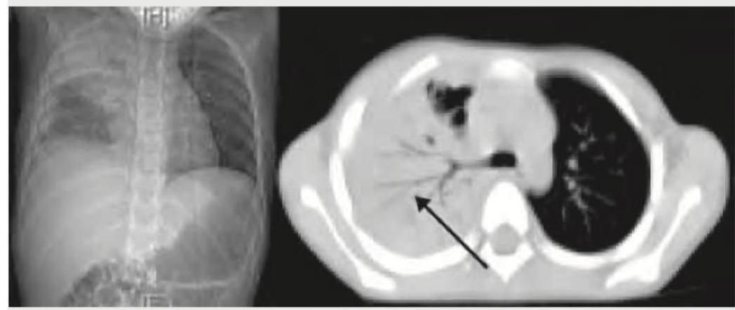
What does this CT chest image show?
A research team develops an AI algorithm using 100,000 CT scans from multiple institutions. The algorithm shows excellent performance (AUC 0.96) but requires extensive computational resources. To deploy it in resource-limited settings, they propose model compression techniques. Evaluate the potential trade-offs and propose the most balanced approach.
A radiology department is evaluating two AI algorithms for fracture detection. Algorithm A has AUC-ROC of 0.95, while Algorithm B has AUC-ROC of 0.92 but provides explainable results showing which image regions influenced its decision. Considering clinical implementation and medicolegal aspects, which statement best evaluates the choice?
A hospital implements an AI algorithm for detecting intracranial hemorrhage on CT scans. The algorithm was trained on data from a different population with different CT scanner protocols. The algorithm shows decreased performance. Which concept explains this phenomenon?
A 55-year-old male presents with chronic cough. A chest X-ray is analyzed by an AI algorithm that reports a 4mm lung nodule in the right upper lobe with 85% confidence. The human radiologist reviews the image but cannot identify the nodule. What is the most appropriate next step?
How does a Generative Adversarial Network (GAN) work in the context of medical image synthesis?
Want unlimited practice?
Get full access to all questions, explanations, and performance tracking.
Scan to download app