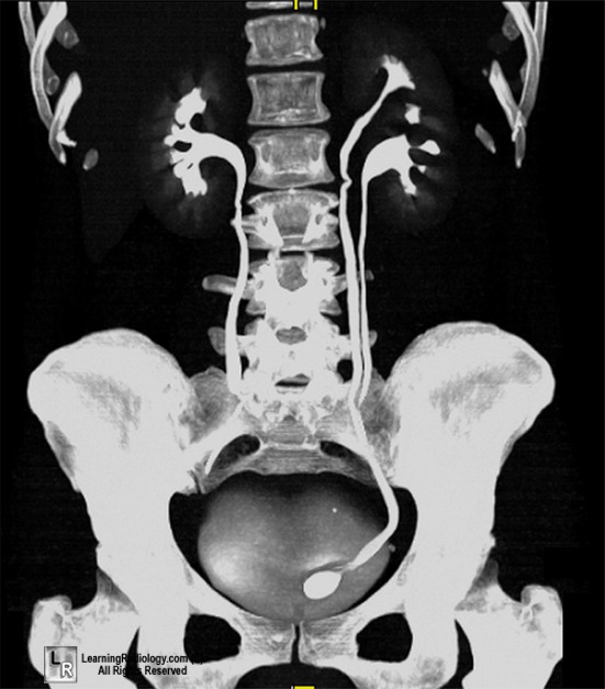
A female patient presented with recurrent urinary tract infections. Imaging shows the following picture. What can be the most probable diagnosis based on the imaging findings?

Cobra head appearance on excretory urography is suggestive of?
Which of the following X-ray findings is associated with Chilaiditi syndrome?
In a patient with a tender and rigid abdomen, what is the expected finding on X-ray?
Rigler's sign is suggestive of?
What is the best investigation for diagnosis and staging of renal cell carcinoma with thrombus extending into the IVC?
"String of beads" appearance on horizontal abdominal view X-ray is suggestive of:
What is the primary use of the Balthazar scoring system?
Hose pipe appearance of intestine is a feature of
The CT severity index in acute pancreatitis is described by:
Explanation: ***Ureterocele (Correct Answer)*** - The image shows a **cystic dilation of the distal ureter** (red arrow) that protrudes into the bladder, which is characteristic of a ureterocele. - Ureteroceles can cause **obstruction and recurrent UTIs** due to stasis and impaired emptying. *Ureteral duplication (Incorrect)* - Ureteral duplication would present as two distinct ureters draining from the same kidney, typically with separate insertions into the bladder or urethra. This is not observed in the image, as there is only one ureter visible from each kidney down to the bladder. - While complete ureteral duplication often features an *ectopic* and *obstructed* upper pole ureter, the imaging here primarily shows a bladder abnormality. *Congenital megaureter (Incorrect)* - A congenital megaureter involves a **diffusely dilated ureter** along its length, without the focal, cystic protrusion into the bladder seen here. - While it can cause UTIs due to stasis, the specific localized dilation inside the bladder strongly points away from this diagnosis. *Urinary calculi (Incorrect)* - Urinary calculi (stones) would appear as **hyperdense opacities within the collecting system** or ureter, which could cause obstruction and UTIs. - The image distinctly shows a **fluid-filled, balloon-like structure** at the ureterovesical junction, not a calcified stone.
Explanation: ***Ureterocele*** - A **cobra head appearance** on excretory urography is a classic sign of a **ureterocele**, which is a cystic dilation of the distal ureter that protrudes into the bladder. - This appearance is due to the dilated ureter appearing like an oval or round filling defect within the bladder lumen, surrounded by a thin radiolucent halo created by the ureteral wall and urine. *Horseshoe kidney* - A horseshoe kidney is characterized by the fusion of the lower poles of the kidneys, causing a **"U" shape** across the midline, often identified by the isthmus. - It does not present with a cobra head appearance but rather a typical anatomical variation of renal position and fusion. *Duplication of renal pelvis* - Duplication of the renal pelvis involves two separate collecting systems draining one kidney, which can be seen as two distinct pelvicalyceal systems. - This condition does not create a cobra head appearance; instead, it shows an abnormal number of collecting systems within a single kidney. *Simple cyst of kidney* - A simple renal cyst typically appears as a **well-defined, anechoic (on ultrasound) or hypodense (on CT) mass** within the kidney parenchyma. - It does not involve the ureter or bladder and thus does not produce a cobra head appearance on urograms.
Explanation: ***Pseudopneumoperitoneum*** - Chilaiditi syndrome is characterized by the **interposition of a loop of colon (usually transverse colon) or, less commonly, small intestine** between the liver and the right hemidiaphragm. - This anatomical variation can mimic **free air under the diaphragm** on an X-ray, leading to the misdiagnosis of pneumoperitoneum. *Pseudopneumothorax* - This term describes the appearance of **air in the pleural space** that is not actually present, which is not associated with Chilaiditi syndrome. - While Chilaiditi syndrome involves misinterpretation of air, it specifically relates to the **abdominal cavity**, not the thoracic cavity. *Pneumothorax* - A **true pneumothorax** is the presence of air in the pleural cavity causing partial or complete lung collapse, which is a significant medical emergency. - It is distinct from Chilaiditi syndrome, which involves **abdominal content displacement** mimicking abdominal free air. *Hydropneumothorax* - This condition involves the presence of both **fluid and air in the pleural cavity**. - It is a pathology of the thoracic cavity and has **no direct association** with the abdominal interposition of bowel loops seen in Chilaiditi syndrome.
Explanation: ***Air under the diaphragm*** - The presence of **free air** (pneumoperitoneum) beneath the diaphragm on an upright abdominal X-ray is a classic sign of **visceral perforation**. - A **tender and rigid abdomen** (peritoneal signs) indicates irritation of the peritoneum, most commonly due to a ruptured hollow viscus. *Blood under the diaphragm* - While blood can accumulate under the diaphragm (e.g., from **trauma** or a ruptured ectopic pregnancy), it typically manifests as a **hemoperitoneum** on imaging. - Blood is **fluid** and would appear as a fluid collection, not free air, on X-ray. *Hazy lung fields* - **Hazy lung fields** suggest conditions like **pulmonary edema**, pneumonia, or acute respiratory distress syndrome (ARDS). - These findings are primarily associated with pulmonary pathology and are not directly indicative of an acute abdominal emergency like perforation. *Prominent vascular markings* - **Prominent vascular markings** often indicate increased blood flow to the lungs or **pulmonary hypertension**. - This finding is unrelated to acute abdominal pain or peritoneal irritation.
Explanation: ***Correct: Pneumoperitoneum*** - **Rigler's sign** (double wall sign) is the visualization of both the **inner (mucosal) and outer (serosal) surfaces** of the bowel wall on an abdominal X-ray. - This occurs when **free intraperitoneal air** outlines both sides of the bowel wall, making it a **pathognomonic sign of pneumoperitoneum**. - Commonly seen in **bowel perforation** from causes like peptic ulcer, trauma, or iatrogenic injury. *Incorrect: Pneumothorax* - Refers to air in the **pleural space** (thoracic cavity), not the peritoneal cavity. - Diagnosed on chest X-ray by the **visceral pleural line** with absent lung markings peripherally. - Completely different anatomical compartment from where Rigler's sign is observed. *Incorrect: Peritonitis* - Represents **inflammation of the peritoneum**, which is a clinical and pathological diagnosis. - While pneumoperitoneum from perforation can **lead to peritonitis**, Rigler's sign specifically indicates the **presence of free air**, not inflammation itself. - Peritonitis has no specific pathognomonic radiological sign like Rigler's. *Incorrect: Hemothorax* - Refers to **blood in the pleural cavity** (thoracic, not abdominal). - Appears as a **pleural effusion** with meniscus sign on chest X-ray. - Unrelated to abdominal radiological findings or free air.
Explanation: ***CT scan*** - **CT scan** with contrast is the gold standard for diagnosing renal cell carcinoma and evaluating the extent of tumor thrombus into the **IVC**. - It provides detailed anatomical information on the tumor, staging, and involvement of adjacent structures. *Angiography* - **Angiography** is an invasive procedure primarily used for mapping the vascular supply of the tumor preoperatively or for embolization, not as a primary diagnostic tool. - It carries risks associated with contrast agents and catheterization and provides less comprehensive detail on tumor extension compared to CT. *Colour doppler imaging* - While useful for detecting blood flow and confirming the presence of a thrombus, **color Doppler imaging** (ultrasound) has limitations in accurately assessing the cranial extent of an IVC thrombus. - Its diagnostic accuracy is highly operator-dependent and less reliable for deep structures like the IVC compared to CT. *IVP* - **Intravenous Pyelogram (IVP)** assesses the urinary tract's structure and function but has limited utility in detecting soft tissue masses like renal cell carcinoma or IVC thrombus. - It involves radiation exposure and contrast material, and has largely been replaced by more advanced imaging techniques like CT and MRI for renal masses.
Explanation: ***Small bowel obstruction*** - A "string of beads" appearance on a horizontal abdominal view X-ray refers to small gas bubbles trapped between the valvulae conniventes in a dilated small bowel loop. - This finding is highly suggestive of **complete small bowel obstruction**, particularly when accompanied by multiple air-fluid levels and dilated bowel loops. *Intussusception* - While it causes obstruction, intussusception usually appears as a **target sign** (doughnut sign) on ultrasound or a **meniscus sign** on barium enema, not a string of beads on plain X-ray. - Plain X-rays may show signs of **bowel obstruction**, but the string of beads is not characteristic. *Sigmoid volvulus* - Sigmoid volvulus is characterized by a **dilated loop of colon** forming an inverted U-shape, often described as a **coffee bean sign** or **omega sign**, on plain X-ray. - This involves the large bowel, and the "string of beads" specifically relates to gas in the small bowel. *Large bowel obstruction* - Large bowel obstruction typically presents with a **dilated colon** proximal to the obstruction and a collapsed distal colon, often with absent or minimal gas in the rectum and sigmoid. - While air-fluid levels can be present, the "string of beads" is a specific sign of gas within dilated small bowel loops, distinguishing it from most large bowel obstructions.
Explanation: ***Acute Pancreatitis*** - The Balthazar score (also known as the **CT Severity Index** for pancreatitis) is primarily used to assess the severity of **acute pancreatitis** based on findings from a **CT scan**. - It evaluates pancreatic inflammation and necrosis, correlating with patient prognosis and the risk of complications. *Acute Appendicitis* - Acute appendicitis is typically diagnosed clinically, often with the help of the **Alvarado score** or imaging like ultrasound/CT, but not the Balthazar score. - The Balthazar score's focus on pancreatic changes is irrelevant to appendiceal inflammation. *Acute Cholecystitis* - Diagnosis of acute cholecystitis is based on clinical signs, lab tests, and imaging (ultrasound showing **gallbladder wall thickening**, pericholecystic fluid, or stones). - The Balthazar scoring system does not apply to the assessment of gallbladder inflammation. *Cholangitis* - Cholangitis is an infection of the bile ducts, diagnosed using the **Tokyo Guidelines**, which consider systemic inflammation, cholestasis, and imaging of biliary obstruction. - The Balthazar score is specific to pancreatic inflammation and does not provide information relevant to cholangitis.
Explanation: ***Crohns disease*** - The **hose pipe appearance** of the intestine on imaging is due to **transmural inflammation** and **strictures**, characteristic of Crohn's disease [1]. - This feature indicates a **narrowed lumen** due to fibrosis, often affecting the small intestine or colon [1]. *Malabsorption syndrome* - This condition is primarily associated with **nutrient absorption issues**, not structural changes in the intestine. - It typically presents with **diarrhea**, **weight loss**, and **malnutrition**, lacking the characteristic imaging findings. *Ulcerative colitis* - Usually presents with **continuous lesions** confined to the colonic mucosa, leading to ulcers and inflammation but not a **hose pipe appearance**. - Symptoms include **bloody diarrhea** and **abdominal pain**, distinctly different from Crohn's disease. *Hirsprung disease* - A congenital condition causing **intestinal obstruction** due to the absence of ganglion cells, leading to **dilated proximal bowel** rather than a hose pipe appearance. - Typically presents in infants with **severe constipation** and **abdominal distension**, unrelated to imaging features seen in Crohn's disease. **References:** [1] Cross SS. Underwood's Pathology: A Clinical Approach. 6th ed. Common Clinical Problems From Alimentary System Disease, pp. 366-367.
Explanation: ***Balthazar score*** - The **Balthazar score** (or CT severity index) is a widely used radiological grading system for assessing the severity of **acute pancreatitis** based on findings on computed tomography (CT) scans. It evaluates both pancreatic inflammation and necrosis. - The Balthazar score helps predict the clinical course and potential complications of pancreatitis by assigning points for **pancreatic inflammation** and the extent of **necrosis**. *Mengini score* - The **Mengini score** is not a recognized CT severity index specifically for acute pancreatitis. - This name is not associated with any established scoring system in gastroenterology. *Chapman score* - The **Chapman score** refers to specific somatic points used in **osteopathic manipulative medicine** for diagnosis and treatment, primarily related to lymphatic system dysfunction. - It has no relevance to the radiological assessment or severity grading of acute pancreatitis. *Napelon score* - The **Napelon score** does not exist as a recognized medical scoring system, particularly in the context of acute pancreatitis or medical imaging. - This name is likely a distractor and not associated with medical practice.
Imaging of Liver
Practice Questions
Biliary Tract Imaging
Practice Questions
Pancreatic Imaging
Practice Questions
Spleen and Lymphatic System
Practice Questions
Gastrointestinal Tract Imaging
Practice Questions
Renal and Urinary Tract Imaging
Practice Questions
Adrenal Imaging
Practice Questions
Female Pelvic Imaging
Practice Questions
Male Pelvic Imaging
Practice Questions
Abdominal Trauma Imaging
Practice Questions
Acute Abdomen Imaging
Practice Questions
Imaging of Peritoneal Cavity and Retroperitoneum
Practice Questions
Get full access to all questions, explanations, and performance tracking.
Start For Free