The most appropriate first-line investigation to be performed in suspected cases of gastric cancer is:
Which imaging modality is considered the best for staging rectal carcinoma?
Which of the following is a direct (primary) sign of obstruction of the urinary tract on a CT scan?
Which of the following HSG findings is most suggestive of genital tuberculosis?
Which of the following is a valid imaging criterion for unresectable carcinoma of the pancreas?
What is the most sensitive investigation for detecting minimal gas in the abdomen?
The 'coffee bean sign' is typically seen in which condition?
Which of the following is a radiological sign of acute pancreatitis on plain radiography?
Depth of gastric carcinomas is assessed by -
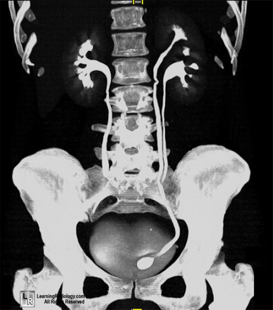
A female patient presented with recurrent urinary tract infections. Imaging shows the following picture. What can be the most probable diagnosis based on the imaging findings?

Explanation: ***Endoscopy*** - **Endoscopy with biopsy** is the most definitive first-line investigation for suspected gastric cancer as it allows direct visualization of the gastric mucosa and tissue sampling for histological confirmation. - It is crucial for early detection, staging, and guiding subsequent management strategies in patients suspected of having gastric malignancy. *Double contrast radiography* - **Double contrast radiography** (barium swallow) can reveal suspicious mucosal abnormalities but is less sensitive and specific than endoscopy for detecting and characterizing early gastric lesions. - It does not allow for **biopsy**, which is essential for definitive diagnosis of cancer. *Plain radiography* - **Plain radiography** (X-ray) has very limited utility in diagnosing gastric cancer as it cannot visualize mucosal lesions or provide detailed information about the gastric wall. - It is primarily used for detecting complications like **perforation** or **obstruction**, rather than primary diagnosis. *CT Scan* - A **CT scan** is valuable for staging gastric cancer after diagnosis, assessing local invasion, lymph node involvement, and distant metastases. - However, it is not the first-line diagnostic modality because it cannot directly visualize early mucosal lesions or provide **histological confirmation** via biopsy.
Explanation: ***MRI*** - **Magnetic Resonance Imaging (MRI)** provides excellent soft tissue contrast, allowing for detailed visualization of the rectal wall layers, mesorectal fascia, and adjacent structures. - This high resolution is crucial for accurate **T-staging (tumor depth of invasion)** and assessment of **nodal involvement**, guiding treatment decisions like neoadjuvant therapy. *CT Scan* - **CT scans** are better for detecting distant metastases (e.g., in the liver or lungs) and assessing vascular involvement, rather than local staging of the primary tumor. - Its soft tissue resolution is inferior to MRI for distinguishing between the different layers of the rectal wall and for precise mesorectal fascia involvement. *TRUS* - **Transrectal ultrasound (TRUS)** is effective for evaluating superficial tumors and their depth of invasion within the rectal wall. - However, it has a limited field of view and is less reliable for assessing mesorectal fascia involvement or metastatic lymph nodes far from the rectal wall. *None of the options* - This option is incorrect because MRI is indeed the preferred imaging modality for precise local staging of rectal carcinoma due to its superior soft tissue resolution. - Accurate staging is critical for determining the appropriate treatment strategy, including the need for neoadjuvant chemoradiotherapy.
Explanation: ***Hydroureter*** - **Hydroureter** (dilation of the ureter) is a **direct/primary sign** of urinary tract obstruction on CT scan. - It represents the physical consequence of upstream blockage and is a **direct visualization** of the obstructed collecting system. - On CT, a dilated ureter proximal to the point of obstruction is the most specific radiological evidence of ureteral obstruction. *Perinephric stranding* - **Perinephric stranding** represents edema in the perinephric fat and is a **secondary/indirect sign** of obstruction. - While commonly seen with acute ureteral obstruction (as part of renal inflammatory response), it is **not a direct sign** of the obstruction itself. - It can also occur with pyelonephritis, trauma, or other inflammatory processes, making it less specific. *Thickening of the lateroconal fascia* - **Lateroconal fascia thickening** is another **secondary/indirect sign** that can accompany urinary tract obstruction. - It reflects inflammatory changes in the retroperitoneal fascial planes adjacent to an obstructed kidney. - Like perinephric stranding, it is a non-specific finding that can occur with various retroperitoneal inflammatory processes. *None of the options* - This option is incorrect because **hydroureter** is a well-established direct sign of urinary tract obstruction on CT scan.
Explanation: ***Beaded tubes*** - **Beading** of the fallopian tubes on hysterosalpingography (HSG) is **the most characteristic finding** of **genital tuberculosis (GTB)**, representing multiple strictures and dilatations. - This appearance is due to the characteristic **granulomatous inflammation** and subsequent fibrosis that occur with tuberculous salpingitis, creating a **"rosary bead" or "string of pearls"** pattern. - This is considered the **most specific HSG sign** for genital TB. *Honeycomb uterus* - A **honeycomb uterus** appearance on HSG shows multiple small filling defects in the endometrial cavity. - This is more commonly associated with **synechiae (Asherman's syndrome)** or advanced **endometrial tuberculosis**, but is not the most suggestive finding. *Golf club tube* - A **golf club tube** appearance refers to a **dilated fallopian tube with a rounded, blunt end** that has lost its fimbrial integrity, seen in **hydrosalpinx**. - While hydrosalpinx can occur due to GTB, this finding is **non-specific** and can result from any chronic salpingitis (PID, post-infectious). *Pipe stem tubes* - **Pipe stem** or **rigid tubes** show straightened, non-dilated fallopian tubes due to fibrosis. - While this can be seen in genital TB, it is **less specific** than the beaded appearance and can occur in other chronic inflammatory conditions.
Explanation: ***Encasement of the superior mesenteric artery*** - **Encasement** (defined as >180-degree circumferential contact) of the **superior mesenteric artery (SMA)** is a definitive **local imaging criterion** for unresectable pancreatic cancer per NCCN guidelines. - This represents the classic teaching point for imaging-based assessment of resectability based on tumor-vessel relationships. - SMA encasement precludes safe surgical resection with negative margins. *Invasion of the duodenal wall* - Duodenal wall invasion is typically managed during **pancreaticoduodenectomy (Whipple procedure)** with en-bloc resection of the duodenum. - Isolated duodenal invasion does not render the tumor unresectable and is expected in most pancreatic head tumors. *Metastatic spread to vertebrae* - While **distant metastases** (including vertebral metastases) absolutely indicate unresectability, they represent **systemic spread** rather than a local imaging criterion for assessing primary tumor resectability. - In clinical practice, the assessment of resectability based on imaging primarily focuses on **local tumor-vessel relationships** (SMA, celiac axis, portal vein, SMV). - Metastatic disease is typically categorized separately under staging rather than resectability criteria based on locoregional anatomy. *Irregular increase in density of omental fat* - Increased omental fat density may suggest **peritoneal carcinomatosis** but is non-specific and not a definitive criterion. - Requires tissue confirmation and is not a standard imaging criterion for determining unresectability.
Explanation: ***CT Scan of the abdomen*** - **CT scans** are highly sensitive for detecting even small amounts of **free air (pneumoperitoneum)** due to their ability to produce cross-sectional images with high spatial resolution missing in conventional X-rays. - It can identify the exact location and quantify the volume of gas, often picking up gas that is not visible on plain radiographs. *Chest X-ray in AP View* - An **AP chest X-ray** is less sensitive for detecting subdiaphragmatic free air compared to an erect chest X-ray or CT scan. - With the patient supine (as implied by AP view if not specified as erect), small amounts of gas tend to spread diffusely rather than collect under the diaphragm. *X-ray abdomen in supine position* - A supine abdominal X-ray is generally the **least sensitive** plain radiograph for detecting free air. - Gas in the abdomen tends to accumulate anteriorly when the patient is supine, making it more difficult to visualize against overlying bowel gas or soft tissues. *X-ray abdomen in erect position* - An **erect abdominal X-ray** or erect chest X-ray is commonly used and more sensitive than a supine view for detecting free air, as gas can rise and collect under the diaphragm. - However, it still requires a sufficient volume of gas to be visible and is less sensitive than a CT scan, especially for very minimal amounts or atypical locations.
Explanation: ***Sigmoid volvulus*** - The **coffee bean sign** is a classic radiographic finding in **sigmoid volvulus**, representing the hugely distended, gas-filled loop of bowel twisting on its mesentery. - This sign is due to the **two limbs of the distended sigmoid colon** converging towards the pelvis, creating a peculiar shape on an abdominal X-ray. *Gastric volvulus* - **Gastric volvulus** involves torsion of the stomach, which can appear as a massively dilated stomach with an air-fluid level or a "double bubble" sign if there is also duodenal obstruction. - It does not typically produce the **coffee bean appearance**, which is characteristic of colonic volvulus. *Hypertrophic pyloric stenosis* - **Hypertrophic pyloric stenosis** is a condition in infants characterized by a thickened pylorus, often leading to non-bilious projectile vomiting. - Imaging typically shows an elongated, narrowed pyloric channel (the "string sign") and a thickened pyloric muscle ("target sign" or "doughnut sign") on ultrasound, not a coffee bean sign. *Achalasia* - **Achalasia** is an esophageal motility disorder where the lower esophageal sphincter fails to relax, leading to food accumulation. - Radiographic findings include a dilated esophagus with a "bird's beak" appearance at the gastroesophageal junction, distinct from the coffee bean sign.
Explanation: ***Sentinel loop sign*** - The **sentinel loop sign** refers to a focally dilated segment of small bowel (usually proximal jejunum) adjacent to an inflamed pancreas due to localized ileus. - This sign is often visible on **plain abdominal radiographs** in cases of acute pancreatitis. *Murphy's sign* - **Murphy's sign** is a clinical finding, not a radiological sign, characterized by inspiratory arrest upon deep palpation of the right upper quadrant. - It is classically associated with **acute cholecystitis**, not acute pancreatitis. *Renal halo sign* - The **renal halo sign** is a radiological finding (typically on CT) describing perinephric fat stranding that can be seen in various renal pathologies, not acute pancreatitis. - It is not a characteristic sign of pancreatic inflammation on plain radiography. *Grey Turner's sign* - **Grey Turner's sign** is a clinical sign characterized by ecchymosis or discoloration of the flanks. - It indicates **retroperitoneal hemorrhage**, which can be a severe complication of acute pancreatitis but is not a radiological sign itself and is not specific to acute pancreatitis on plain X-ray.
Explanation: ***Endoluminal ultrasound*** - **Endoluminal ultrasound (EUS)** utilizes a small ultrasound transducer at the tip of an endoscope to provide high-resolution images of the **gastric wall layers**, making it ideal for assessing the **depth of tumor invasion**. - EUS can accurately stage the **T-stage (tumor invasion depth)** and detect **regional lymph node involvement** (N-stage), which are crucial for treatment planning in gastric carcinoma. *Abdominal ultrasound* - **Abdominal ultrasound** has limited ability to visualize the fine layers of the gastric wall and is primarily used for detecting **larger masses**, **liver metastases**, or **ascites**. - It is not precise enough to determine the **depth of tumor invasion** in gastric carcinoma. *Barium meal* - A **barium meal** (upper GI series) is a radiographic study that visualizes the lumen of the esophagus, stomach, and duodenum, primarily detecting **mucosal abnormalities** or **filling defects**. - While it can identify the presence of a tumor, it provides no information on the **depth of penetration** into the gastric wall. *Laparoscopy* - **Laparoscopy** is a surgical procedure that allows for direct visual inspection of the peritoneal cavity and offers the best method for detecting **peritoneal carcinomatosis** or **distant metastases**. - While it can confirm the presence of a tumor and help in operative planning, it does not provide detailed information about the **depth of intramural invasion** of the gastric wall itself, which is best assessed by EUS.
Explanation: ***Ureterocele (Correct Answer)*** - The image shows a **cystic dilation of the distal ureter** (red arrow) that protrudes into the bladder, which is characteristic of a ureterocele. - Ureteroceles can cause **obstruction and recurrent UTIs** due to stasis and impaired emptying. *Ureteral duplication (Incorrect)* - Ureteral duplication would present as two distinct ureters draining from the same kidney, typically with separate insertions into the bladder or urethra. This is not observed in the image, as there is only one ureter visible from each kidney down to the bladder. - While complete ureteral duplication often features an *ectopic* and *obstructed* upper pole ureter, the imaging here primarily shows a bladder abnormality. *Congenital megaureter (Incorrect)* - A congenital megaureter involves a **diffusely dilated ureter** along its length, without the focal, cystic protrusion into the bladder seen here. - While it can cause UTIs due to stasis, the specific localized dilation inside the bladder strongly points away from this diagnosis. *Urinary calculi (Incorrect)* - Urinary calculi (stones) would appear as **hyperdense opacities within the collecting system** or ureter, which could cause obstruction and UTIs. - The image distinctly shows a **fluid-filled, balloon-like structure** at the ureterovesical junction, not a calcified stone.
Imaging of Liver
Practice Questions
Biliary Tract Imaging
Practice Questions
Pancreatic Imaging
Practice Questions
Spleen and Lymphatic System
Practice Questions
Gastrointestinal Tract Imaging
Practice Questions
Renal and Urinary Tract Imaging
Practice Questions
Adrenal Imaging
Practice Questions
Female Pelvic Imaging
Practice Questions
Male Pelvic Imaging
Practice Questions
Abdominal Trauma Imaging
Practice Questions
Acute Abdomen Imaging
Practice Questions
Imaging of Peritoneal Cavity and Retroperitoneum
Practice Questions
Get full access to all questions, explanations, and performance tracking.
Start For Free