
Abdominal and Pelvic Radiology — MCQs

On this page
What does the following radiograph from a double contrast esophagram represent?
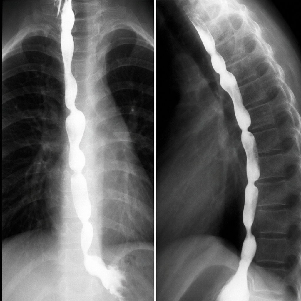
Chain of lakes appearance is seen in?
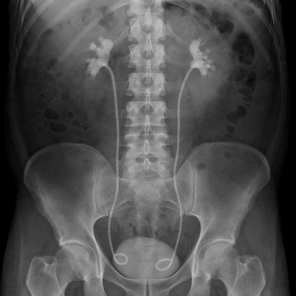
What are the bilateral radiopaque structures extending from the renal pelvis to the bladder seen in this abdominal X-ray?
Percentage of renal stones which are radio-opaque:
Which of the following is a direct (primary) sign of obstruction of the urinary tract on a CT scan?
Investigation of choice for detecting hepatic metastasis from stomach cancer is
Which of the following HSG findings is most suggestive of genital tuberculosis?
Which imaging modality is considered the best for staging rectal carcinoma?
Which of the following is a radiological sign of acute pancreatitis on plain radiography?
The 'coffee bean sign' is typically seen in which condition?
Practice by Chapter
Imaging of Liver
Practice Questions
Biliary Tract Imaging
Practice Questions
Pancreatic Imaging
Practice Questions
Spleen and Lymphatic System
Practice Questions
Gastrointestinal Tract Imaging
Practice Questions
Renal and Urinary Tract Imaging
Practice Questions
Adrenal Imaging
Practice Questions
Female Pelvic Imaging
Practice Questions
Male Pelvic Imaging
Practice Questions
Abdominal Trauma Imaging
Practice Questions
Acute Abdomen Imaging
Practice Questions
Imaging of Peritoneal Cavity and Retroperitoneum
Practice Questions
Want unlimited practice?
Get full access to all questions, explanations, and performance tracking.
Scan to download app